Takeaways
- Galloping and specialized gaits place immense stress on the equine forelimb, leading to carpal hyperextension (backward slanting of the cannon bone) in a significant portion of competing horses.
- Muscle fatigue, impact forces (such as landing over jumps or momentum from weighted boots) and poor conformation are primary drivers of carpal hyperextension, increasing strain on tendons and the suspensory apparatus.
- Practices like using toe grabs higher than 2 mm, long-toe/low-heel trimming, or prolonged wearing of weighted “performance boots” contribute to joint derangement, coffin bone rotation, and a heightened risk of catastrophic injuries.
Click here for Part 1: 13 Rules of Equine Movement
Click here for Part 2: The Equine Walk: “Mother of All Gaits”
Click here for Part 3: How the Trot & Pseudo-Trot Affect the Equine Hoof
Click here for Part 4: The Spectrum of Ambling Horse Gaits
Click here for Part 5: Disarticulating the Horse’s Canter
Click here for Part 6: The Gallop: Horses at Speed
The very fact that a horse is called upon to gallop implies a high level of athletic demand, higher than that typically required by the walk, amble or trot gaits that we have previously reviewed in this series. In our last installment, we detailed the biomechanics and stride characteristics of galloping. This prepares us to now take a closer view of some possible side effects of galloping, especially as they relate to the hoof but also to the horse’s carpal joint or “knee.” In this article, I also compare effects of “extreme” forms of amble and canter that similarly affect the carpus and surrounding soft tissues.
Finish-line photos of galloping races are easy to find online. However, typically, the high-speed cameras used, while accurately capturing the position of the finishers’ noses, at the same time distort the limbs, and I have therefore not used them to produce this analysis. Instead, I preferred news photographs, usually taken at closer range and with the camera lens on the same level as the horse.
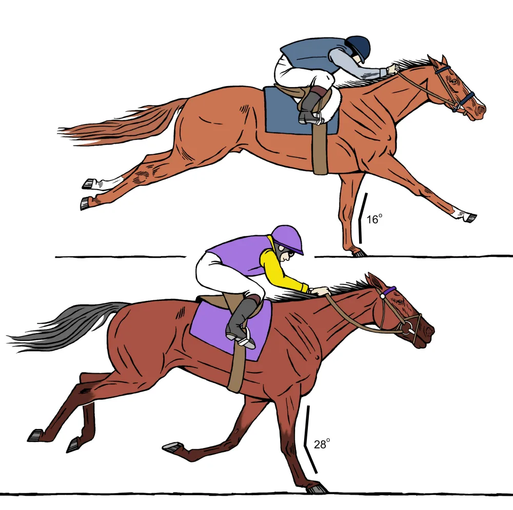
A survey of 500 news photos taken either during fast workouts or during a race forms the basis of this analysis. Race distances varied from two-furlong (quarter mile) contests to “classic distances” of up to 1½ miles. What the survey looked for was carpal hyperextension in the weight-bearing limb during the fourth “beat” of gallop. In this beat, the horse’s weight is supported by a single forelimb, with none of the other limbs in contact with the ground. Carpal hyperextension occurs when the cannon bone of this limb, which should be vertical during weight-bearing (Figure 1A), instead slants backward to any degree (Figure 1B).
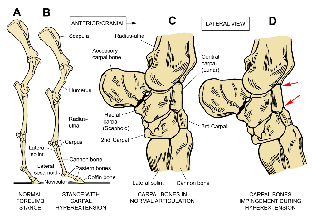
Figure 2. Bony anatomy of the forelimb. A, whole limb in normal stance; B, stance with hyperextension of the carpus. C, close-up of the carpal joint in lateral view, showing normal articulation of the bones that comprise the joint. D, showing articulation during hyperextension of the joint. Red arrows mark areas where hyperextension forces the anterior edges of the central and 3rd carpal bones to impinge upon each other, increasing the likelihood of chip fractures.
Most racehorse photos reviewed — 55% in round numbers — do not show hyperextension of the carpus. A significant minority (40%), however, showed from 4-12 degrees of hyperextension; 5% showed from 1-4 degrees. The alignment of equine forelimb bones in normal relationship is compared in Figure 2 with the articulation when the carpus is hyperextended, which causes the anterior edges of the bones to impinge upon one another (red arrows).
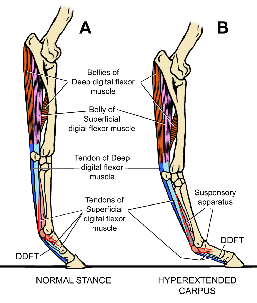
Figure 3. The effect of carpal hyperextension is to increase the tension in the long, strap-like tissues that run down the back of the limb, namely, the deep and superficial digital flexor muscles and their associated long tendons. The suspensory apparatus is also tensioned since carpal hyperextension forces accompany extension of the fetlock joint.
The consequences for soft tissues are made clear in Figure 3. This is yet another reason for the farrier to be aware that there is no such thing as a “tendon,” i.e., a tissue that exists independently of association with anything else. All tendons are part of a muscle; they are extensions of the very cells that make up the belly of the muscle (please see “Connecting Tendon to Bone” in the December 2019 issue of American Farriers Journal). The farrier must realize the “tendons” that one can palpate on the back of the cannon bone extend upward far past the knee to attachment points near the elbow.
In a normal horse, there is complete continuity of these long, strap-like muscles from the arm all the way down to the bottom of the coffin bone. If the farrier’s mental picture of horse anatomy above the knee is a blank, he or she will be unable to fully understand forelimb function (please see “Do You Know the Nuts and Bolts of the “Orthopedic Trim?” in the July/August 2020 issue of AFJ, and “Manipulating the Equine Forelimb with Farriery” in the December 2020 issue of AFJ).
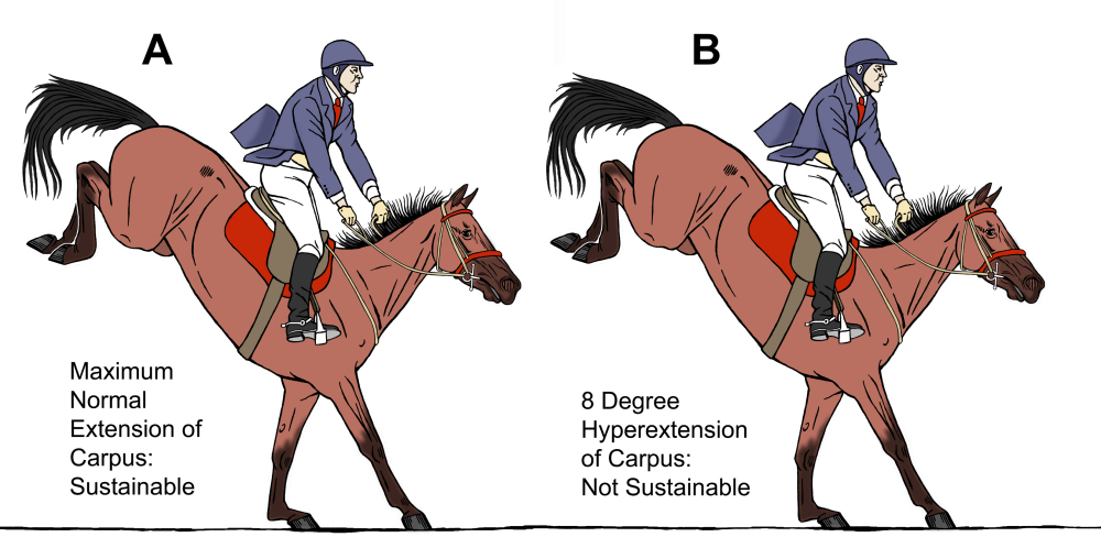
Figure 4. Sustainable and non-sustainable extension of the carpus in a jumper. Horse A shows less than 2 degrees of carpal hyperextension; horse B shows 8 degrees.
Carpal hyperextension can also be observed to occur in competitions other than racing, where galloping is part of the athletic requirement (Figure 4). A survey of 415 photographs of jumpers and 3-day Eventers again shows that the majority (85%) do not show carpal extension as defined above, but that 15% of photos do show it occurring. In these horses, carpal hyperextension does not occur “on the flat,” but as a consequence of landing over the jump.
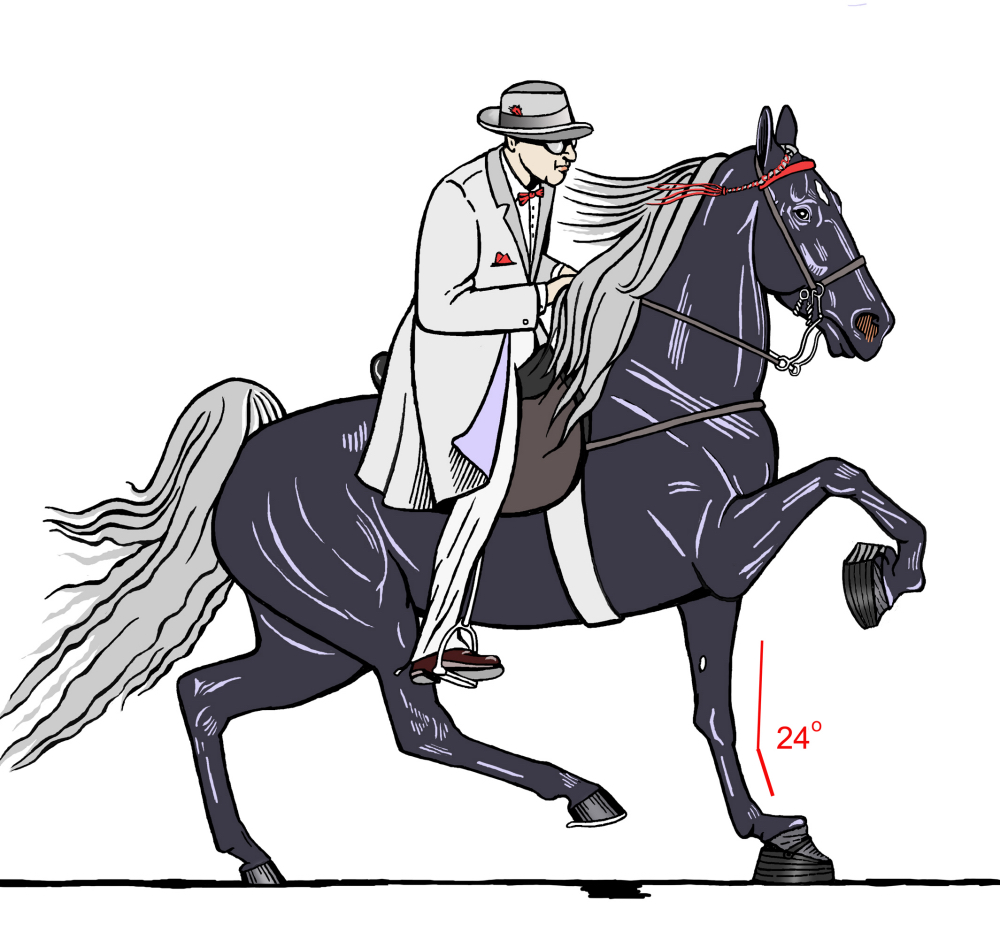
Figure 5. “Big lick” Tennessee Walking Horse wearing weighted “performance boots” held on by nails and bands (he also wears long trailers on the hind hoofs, another bad idea but not covered in this article). He is performing the running walk gait. The degree of carpal hyperextension is about 8 degrees.
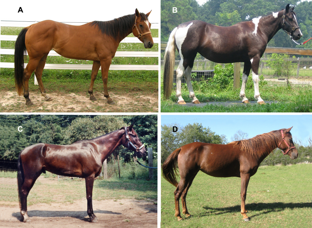
Figure 7. Poor forelimb conformation. A, B, examples of congenital calf-knee. C, D, examples showing insubstantial “bone,” i.e., on the order of 6½ inches or less of bone-tendon circumference per 1,000 lbs. of weight. Both these horses also show small, round joints at the level of both carpus and fetlock.
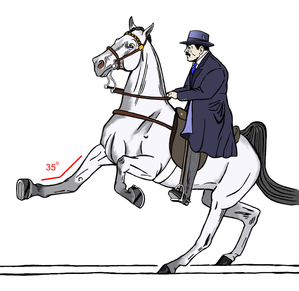
Figure 6. “Big lick” Tennessee Walking Horse wearing weighted “performance boots” performing the canter. The degree of carpal hyperextension is extreme, at about 19 degrees. Note that since the limb is not weight-bearing, this is entirely due to jerk as a result of momentum rather than to compression.
Causes of Carpal HyperextensionThe obvious culprit for carpal hyperextension is faulty conformation. The carpus is hyperextended in horses that have “calf-kneed” forelimb conformation (Figures 7A & 7B; Figure 8B) before the animal even moves. It’s easily hyperextended in horses with weedy conformation or small, round joints (Figures 7C & 7D). Throughout their lives, these horses are going to need trimming and shoeing that speeds breakover and supports the caudal portion of the foot. Such horses are not uncommon in the general population, but they are exceedingly rare at the racetrack or among jumpers or eventers, since it has long been known that such animals have difficulty standing up to the athletic demand due to chip fractures to the carpal bones and proximal cannon, suspensory tears, bowed tendons and navicular changes or “caudal foot syndrome.”
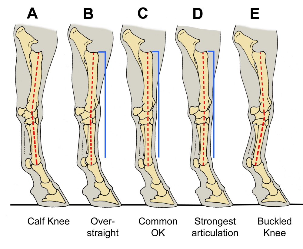
Figure 8. The spectrum of carpal articulation as viewed from the side. The ideal is not to have the dotted red line vertical, but rather to have it deflect slightly to the front to compensate for the fact that the radius-ulna bone is curved. Thus, example C, in which the line is vertical, is “OK,” but example D is the strongest and best conformation. Example A is frankly calf-kneed, i.e., hyperextended in stance; example B is “over-straight,” a conformation commonly seen in which there is a barely detectable degree of static hyperextension. Breeding two “over-straight” horses together is dangerous because it may result in a doubling-up to produce a horse that is frankly calf-kneed.
A famous example of a calf-kneed jumper was the Trakhener stallion Abdullah (gold medalist at the 1984 Los Angeles Summer Olympic Games). Abdullah was extensively bred after winning that and other championships, and without a doubt, passed on his conformation defect along with his jumping ability. Nonetheless, we generally cannot look to conformation as a cause for the carpal hyperextension observed in horses engaged in high-demand athletic activity. We will see below how correct conformation in horses with good bone substance and correct carpal articulations (Figure 8D) can, on the other hand, serve to protect from carpal hyperextension.
In racehorses, carpal hyperextension is most observed as the horses near the finish line. This implicates muscle fatigue as a likely cause. Fatigue causes laxity (inability to contract) in affected muscles and effectively lengthens their tendons. As Figure 3 shows, laxity in the superficial and/or deep digital flexor muscles permits the carpus to sag backwards into the hyperextended position. There is a simultaneous effect on the suspensory apparatus as well, since carpal hyperextension when the hoof is in contact with the ground causes extension of the fetlock joint.
Can muscle fatigue also be a cause of the carpal hyperextension seen in jumpers (Figure 4)? Here, the hyperextension is caught by the camera upon landing over jumps. In the absence of myography carried out on test jumpers connected by Bluetooth to a computer, it’s not possible to rule out muscle fatigue as a cause. However, it’s also reasonable to assume that the impact of landing itself is a factor — my veterinary mentor Dr. Matthew Mackay-Smith, who not only had 50 years of experience in practice but who participated in many jumping events, used to refer to open jumper contests as “falling out of second-story windows.”
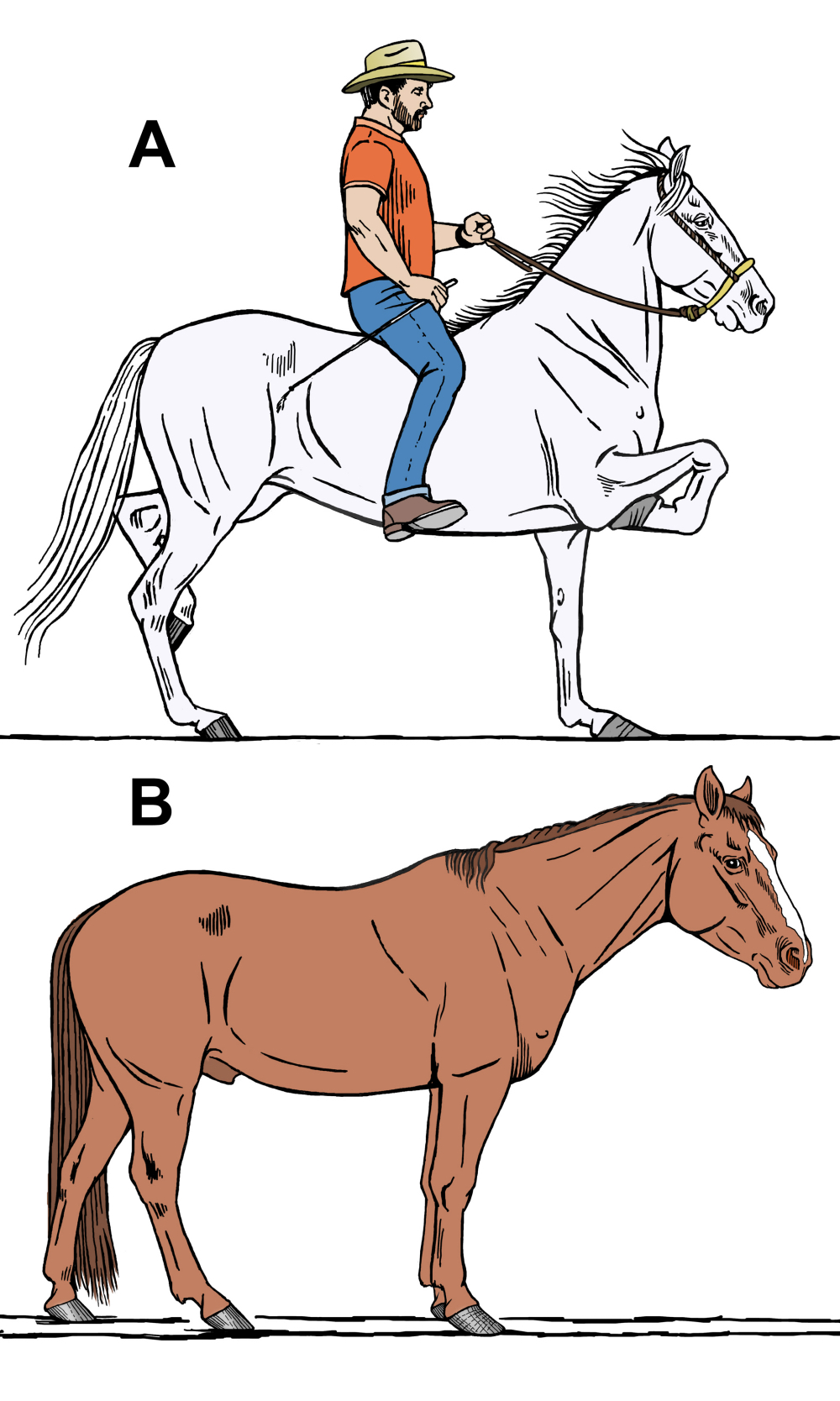
Figure 9. A, a champion Costa Rican mare moving at a trot and showing extremely high, tight forelimb folding. Note the long-toe, low-heel evident in both forefeet. B, a pleasure-riding Quarter Horse that acquired long-toe, low-heel fore hooves due to his owner’s adherence to false ideas of how to trim a horse’s feet. Both these horses have excellent bone substance, at least 8 inches of bone-tendon circumference per 1,000 lbs. of weight, which protects them from hyperextension of the carpus.
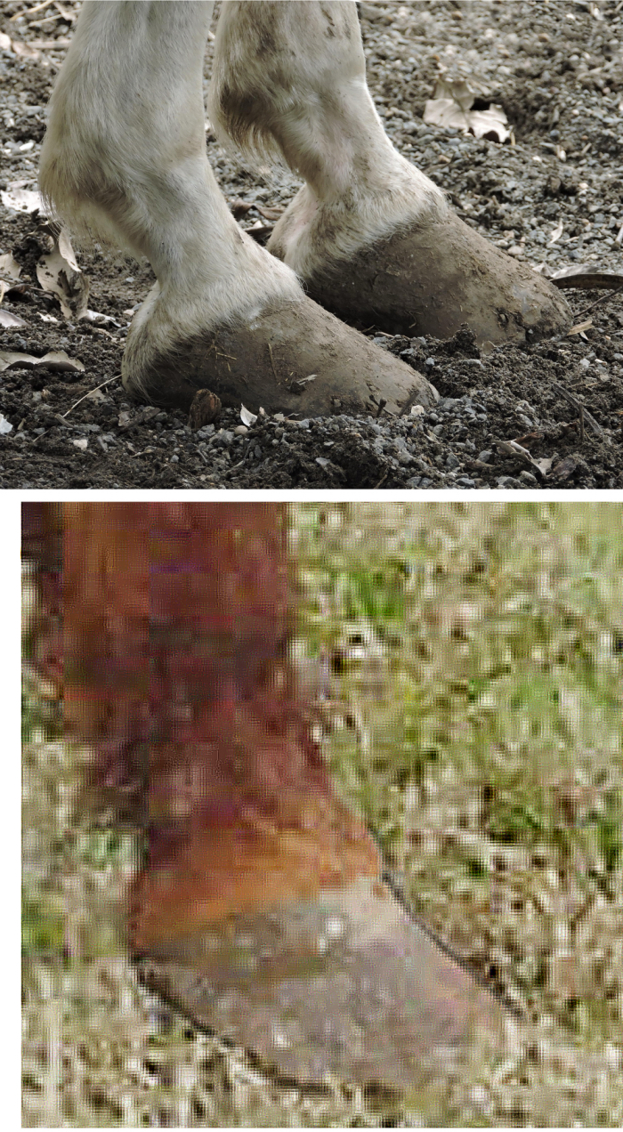
Figure 10. Close-up views of the forehooves of the horses shown in Figure 9. Above, the Costa Rican mare’s forefeet. Below, the forefoot of the Quarter Horse shown in Figure 9B. Long-toe, low-heel conformation has to be developed by repeated mis-trimming; no horse is born with such hooves, nor would any horse develop such a hoof if not mis-trimmed over a period of months or years.
Yet another cause of carpal hyperextension is the “long toe, low heel” configuration of the fore hooves. In Figures 9 & 10, I have chosen two examples that came about neither by accident nor from ignorance of how to trim correctly, but by the deliberate effort of the owner/trainer. In Figures 9A & 10A, the desire was to force extremely high, tight forelimb folding. The mare pictured won a national championship in Costa Rica because of this. By contrast, Figures 9B & 10B show a Quarter Horse — not a breed in which high forelimb “action” is usually wanted. Here instead, the owner was trying to save money by not paying a professional to trim her horse’s feet. Following the erroneous idea that the toe of the hoof should be a direct continuation of the slope of the pasterns, after 2 years she had created almost the same foot as the one deliberately made by the Costa Rican estancionero riding the grey in Figure 9A. But notice, neither the Quarter Horse gelding nor the Costa Rican mare shows any but the most subtle degree of carpal hyperextension, despite the gross increase in leverage that long-toed hooves exert upon the carpus. The reason for this is conformation — both horses have exceptionally substantial “bone,” meaning that the cannon-tendon circumference as measured just below the knee is on the order of 8 inches per 1,000 lbs. of body weight. This is the ideal for riding horses, yet most do not come up to this standard. Both also have broad knees that are shield-shaped in front view, and that in side view articulate correctly both above to the radius-ulna and below to the cannon bone (Figure 8). Excellent conformation is protective; the unfortunate part is that protection ever be needed.Finally, we can mention momentum as a cause for carpal hyperextension. Compare the Tennessee Walking Horse in Figure 6 to the one in Figure 5 and, for that matter, to all the other horses shown in this article. Figure 6 is the only animal showing carpal hyperextension in a limb that’s in the air and not in contact with the ground. It cannot, therefore, be due to impact, although muscle fatigue may play a role. The major cause is the weighted boot. A “slug” is the unit used by physicists to quantify the force of a blow, such as from a wrecking ball swinging by a chain or a roundhouse punch by a boxer. A horse forelimb measuring 4 feet long, swung like a pendulum and bearing a shoe weighing 1 lb., has a potential impact force of 3.19 slug-pounds. The same forelimb bearing a shoe or boot weighing 5 lbs. swings with approximately five times the amount of force in slug-pounds. If the limb does not impact anything, the effect translates into jerk exerted by the heavy distal limb upon the carpus and indeed upon all forelimb joints above the coffin joint.
Given these figures, the derangement of normal carpal articulation evident in Figure 6 cannot be too surprising. What’s surprising is the ability of the animal to survive gross carpal hyperextension — yet “big lick” Tennessee Walkers sometimes have careers several years long. I attribute this, once again, to the protective effect of excellent standing conformation — good to excellent bone substance plus correct articulation of all the joints within and surrounding the carpus (Figure 8).
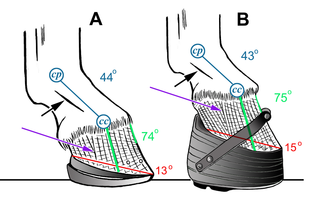
Figure 11. Both images shown have been redrawn directly from photographs. A, a horse undergoing hoof rehabilitation/lameness therapy that has been fitted with wedge pads and a wedged aluminum shoe. B, a Tennessee Walking Horse wearing a “performance boot” held on by nails and a metal band. As the figures in the image show, there is not a lot of difference in terms of function: in both cases, the wedge forces positive-plane rotation of the coffin bone. But because the Walking Horse will live in his “performance boots” continuously for up to several years, they create very deleterious effects which the temporary therapeutic use of wedge pads and wedged shoes mostly avoid. Abbreviations: “cp” is center of rotation of the fetlock joint; “cc” is center of rotation of the coffin joint. The line defining the center axis of the coffin bone (green) matches the angle of the toe above the deepest part of the dish. The red line marks the slope from the buttress to the center of the toe. The blue line is the pastern angle, which is abnormally low due to the wedging. The purple arrow marks the zone of impingement, where the tubules of the heels and quarters (which have run forward) crash into the tubules of the toe. The black arrow indicates the edge of the extensor process of the suspensory apparatus, which comes under tension to the degree that the heels of the hoof are wedged up.
Farrier ResponseA few facts that emerged from questions raised by this survey of carpal hyperextension deserve to be mentioned. First, there’s not a lot of difference in measurable parameters or in terms of breakover between the wedged and rolled therapeutic setup in Figure 11A and the stacks and bands shown in Figure 11B.
To be sure, weights hidden inside the stack or boot, and the height of the boot itself, will ensure that the horse wearing the rig in 11B will move and respond differently than the horse in 11A. It’s important to note that while their heels are wedged up, both animals show positive-plane rotation of the coffin bone and the dished toe that goes with it. Positive-plane rotation forces the pasterns into a more horizontal orientation; the strain this imposes upon the suspensory apparatus can be appreciated by noticing that the edge of the suspensory extensor process becomes prominent — visible and easily palpable (black arrow). Both hooves show a zone of impingement, where the tubules of the heels and quarters that orient at a low angle because the buttresses have run forward, crashing into the tubules of the toe (purple arrow).
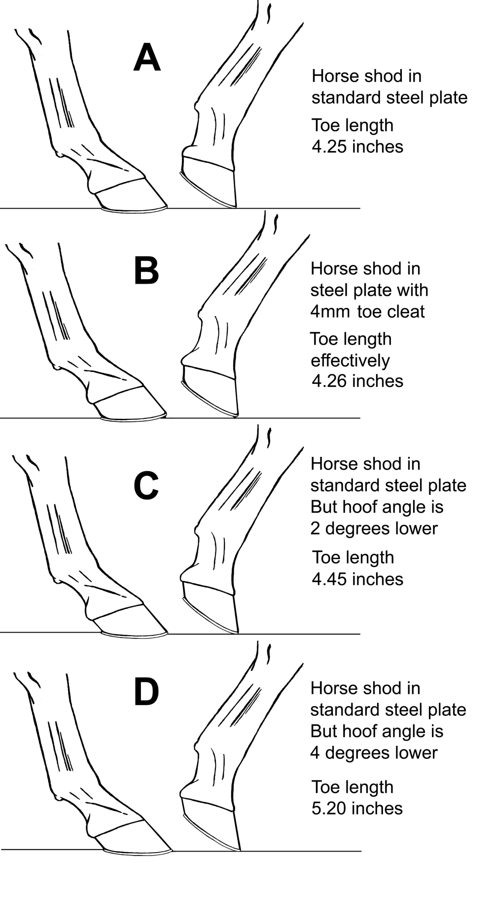
Figure 12. Precision drafting comparing the effect on the speed of breakover on a horse shod in standard plates (A), shod with a 4 mm toe cleat (B), shod with standard plates but with the hoof angle lowered by 2 degrees (C) and by 4 degrees (D). Long-toe, low-heel appears to be somewhat harder on the horse, but toe grabs are not far behind, and many studies have demonstrated their deleterious effects.
The effects of toe grabs — commonly used at the racetrack — vs. long toe, low heel are summarized in Figure 12. Here, I’ve had to do some precision drafting, because in the first place the effect of a 2 mm toe cleat, which is the highest cleat now legal at most American racetracks, is so small that they do not show on a figure drawn to 25% scale. I show a 4 mm cleat in Figure 12B as the smallest one whose effect on breakover can be measured on this type of diagram. Compare Figures 12A, B, C & D and you will find very little difference between a 4 mm toe grab and a 2-degree or even a 4-degree lowering of the toe angle.
Small numbers, however, often translate into big real-world effects. The 2 mm toe grab rule was instituted in response to numerous studies which demonstrate that cleats have consistent harmful effects on equine joints, muscle, tendons and suspensory apparatus (some of the best studies are cited in the references at the end of this article). One important study puts it very succinctly: “The odds of catastrophic musculoskeletal injury in racehorses shod with toe grabs on front shoes was 1.5 times the odds in horses shod without toe grabs — after controlling for age, sex, number of days since last race and cumulative number of furlongs. …The risk increased with increasing toe grab height, with 3.5- and 15.6-times increased risk for fatal musculoskeletal injury and suspensory apparatus failure, respectively, for horses shod with a regular height (2 or 4 mm toe grab) compared with horses shod without a toe grab” (Dahl et. al., 2016).
As we have seen, long toe-low heel delays breakover in the same way the breakover of a person wearing clown shoes or swim fins would also be delayed. It also forces tighter flexion of the carpus and may produce higher “action.” “Low heels and long toe are common means of trimming the foot due to the erroneous belief that it facilitates quicker breakover and thus increases speed” (Hagen et al. 2021). “The greater effort required to rotate the hoof around the long [toe] fulcrum is associated with increased tensile stress on the [deep digital flexor tendon], and a greater compressive force on the navicular bone” (Clayton, 1989). “Increased length of toe increased the odds of sustaining a carpal chip fracture … as the hoof length ratio increased, the odds of sustaining carpal osteochondral fractures increased” (McIlwraith et. al., 2003).
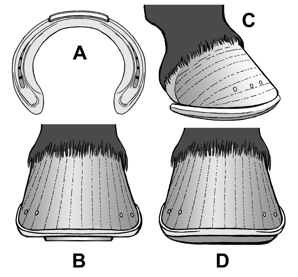
Figure 13. A horseshoe with 4 mm toe cleat (A) and fore hoof shod with this type of shoe in front view (B). The effect of this setup is to delay breakover, increase leverage exerted upon the limb by the toe, and increase tension and strain on the flexor and suspensory apparatus. The exact opposite is the slightly rockered shoe with a rolled toe seen in lateral (C) and frontal view (D). The effect of this setup is to speed breakover and decrease strain upon the flexor tendons and suspensory apparatus. Shoes such as A and B are used for one purpose: to enhance performance; shoes such as C, D are used primarily for therapeutic and rehabilitative purposes on horses already showing signs of lameness.
The difference between a horse shod with a 4 mm toe cleat (Figure 13A & 13B) and a horse shod with a rockered and rolled-toe shoe (Figure 13C & 13D) may not look like much, but these essentially therapeutic designs are nonetheless crucial to the welfare of the animal. When the mere fact of galloping is combined with athletic demand likely to produce muscle fatigue, adding any factor that increases the likelihood of carpal hyperextension is both unreasonable and unjustifiable. The current rule allowing cleats of only 2 mm height is a good one as it favors the horse’s welfare (not to mention avoiding catastrophic injuries that inevitably create bad press for the racing industry).
As to weighted boots, these almost always wedge up the heel relative to the toe. Every horse I have examined that was currently in, or that had recently been taken out of “performance boots” as used in Tennessee Walker competition, or “stacks and bands” as used particularly in Fine Harness American Saddlebreds, showed clear signs of rotation of the coffin bone, including grossly run-under heels, long toes with wide separation of the white line around the toe and founder-type compression rings that converge at the deepest point of the dish in the toe.
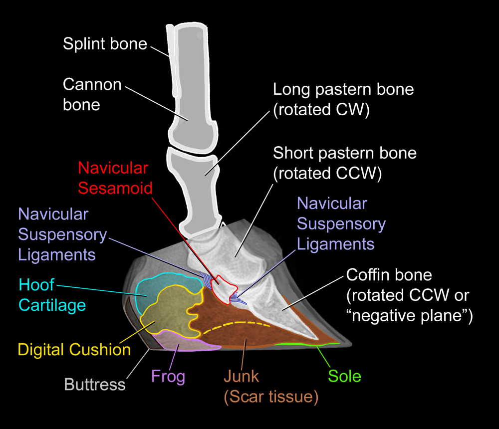
Figure 14. Once the Walking Horse or fine-harness Saddlebred is taken out of wedged shoes, it is crucial for the veterinarian and farrier to recognize that the positive-plane rotation imposed by the wedging during the years when the horse wore the appliance is reversed to negative-plane rotation the moment he is taken out of the appliance. The strain this imposes on all hoof structures is enormous. Tension on the deep digital flexor tendon may be so great that it tears the interlaminar connection by pulling the coffin bone back into positive-plane rotation. Be aware that this is what radiographs taken to guide rehabilitation may initially show. The articulations between all distal joints will likewise be deranged, so that the object of rehabilitation will primarily be to re-establish normal articulations first at the coffin joint and then at every joint higher up.
Radiographs such as the one presented in Figure 14 demonstrate derangement of the articulation of every joint of the distal limb. Thus, while wedging up the heels to relieve tension on the flexor muscle tendons is certainly a viable therapeutic option (Figures 13C & 13D), wedging up the heels when the only reason for doing so is to enhance performance shortens the horse’s competitive career while being seriously detrimental to his long-term health and soundness — sometimes in my experience to the point of being fatal.In short, a horse that’s going to be retired from competition and has been in “performance boots” or wedged stacks for any length of time beyond 2 days cannot simply have its shoes/appliances removed, trim the toe back, and be OK. The radiograph in Figure 14 is a picture of such a horse. To prevent the cascade of deleterious effects that are consequent upon the sudden switch from positive-plane to negative-plane rotation of the coffin bone that occurs when wedged appliances are removed, the horse must be put back into some kind of wedged boot. Over a period of months, the height of the wedge can cautiously be reduced, little by little. How much to reduce and when must be guided by radiographs taken just before re-packing the boots. Re-packing and re-evaluation may need to be done at first as often as once per week, later at monthly intervals.
The several causes for carpal hyperextension may largely be out of the farrier’s control. What’s always under his or her control, however, is which sector of the industry to work in — in other words, to be or not to be the one who puts the “performance boots” on the Walking Horse; or to be or not to be the one who puts shoes with cleats or “toe grabs” higher than 2 mm on the racehorse. Alternatively, the farrier can be the one who applies therapeutic shoes or appliances to the feet of horses that have been crippled by shoeing for “extreme” performance.
I have recently been pleased and impressed to notice that Simon Curtis, PhD, an International Horseshoeing Hall of Famer and frequent keynote speaker at the International Hoof Care Summit, has produced several volumes concerning rehabilitation of the equine hoof and distal limb. This has long been needed, and I applaud any farrier who obtains and studies Curtis’ books.
Resources for Further Reading
- Balch OK, Helman RG & Collier M.A. (2001) Risk factors for catastrophic musculoskeletal injuries in Oklahoma racehorses. Proceedings of the Annual Convention of the AAEP 47:334-338.
- Clayton H. (1989) Toe grabs. Journal of Equine Veterinary Science 9(6):334.
- Dahl VE, Hitchens PL & Stover SM. (2016) Effects of racetrack surface and nail placement on movement between heels of the hoof and horseshoes of racehorses. American Journal of Veterinary Research 77(9):983-990.
- Hagen J, Bos R, Brouwer J, Lux S & Jung FT. (2021) Influence of trimming, hoof angle and shoeing on breakover duration in sound horses examined with hoof-mounted inertial sensors. Veterinary Record 189 (4): 1-9.
- Kane AJ, et al. (1996) Horseshoe characteristics as possible risk factors for fatal musculoskeletal injuries of Thoroughbred racehorses. American Journal of Veterinary Research 57(8): 1147-1152.
- McIlwraith CW, Anderson TM & Sanschi EM. (2003) Conformation and musculoskeletal problems in the racehorse. Clinical Techniques in Equine Practice 2(4):339-347.
- Stover, S.M. 2003. The epidemiology of Thoroughbred racehorse injuries. Clinical Techniques in Equine Practice 2(4):312-322.
- Werner HW. (2012) The Implications of Therapeutic Farriery in Equine Practice. Veterinary Clinics: Equine 28:263-281.