Coronitis and canker may be distinct hoof problems, but they’re often linked – and both can cause lasting damage if not addressed early. Recognizing the signs, understanding their connection, and taking prompt action with coordinated care from a veterinarian, farrier, and horse owner are key to successful treatment.
Link Between Coronitis & Canker
While early texts may not explicitly connect coronitis and canker, the progression from inflammation of the coronary band (coronitis) to more severe hoof conditions like canker was understood in practice and discussed in H. Caulton Reeks’ 1906 Diseases of the Horse’s Foot.
Contemporary veterinary literature acknowledges that chronic inflammation or injury to the coronary band can predispose horses to canker, especially when combined with environmental factors. This understanding reflects a continuity in recognizing the relationship between these conditions over the past century.
Without going in depth, canker is defined as an anaerobic (grows in the absence of oxygen) infection in the superficial epithelium of the hoof or chronic proliferative pododermatitis of yet unknown etiology (Menzies-Gow et al. 2002). The condition is often characterized by abnormal tissue growth, often with a foul odor, and is challenging to treat. It often starts in the frog and potentially spreads to other parts of the hoof (Menzies-Gow et al. 2002).
Like canker, coronitis and coronitis associated with canker does not affect the herd, only certain individual horses.
Signs & Causes of Coronitis
In addition to the coronary band being involved in coronitis, the ergots and chestnuts also can be involved. They can become enlarged, crusty and bleed (Williams 1882; Mahon 1900; Reeks 1918).
In a minority of advanced and long-standing coronitis cases, hoof cracks can occur. This appears to be a weakening of the hoof wall due to coronitis, but the mechanism and why this happens are unknown (Williams 1882; Mahon 1900; Reeks 1918; Reilly 2024; Buff & Reilly 2025). This was thought to be an issue with only draft or draft crosses; however, it is well-documented in many breeds.
The clinical signs include flaking/scaling, crusting, exudate or ulceration of the hoof and the coronary band area.
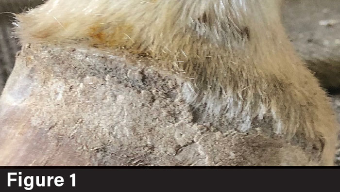
The coronary band swells, causing the hair to stand up or lift away from the hoof wall (Figure 1). The hair can be nearly parallel to the ground. Wart-like scaling of the periople can occur. The hoof wall loses its shiny periople appearance and becomes increasingly flaky.
Some horses are lame and initially mistaken for abscessing or acute laminitis. It can be painful to the touch and the horse can be lame. The coronary band is often warmer than normal (Williams 1882; Merriam-Webster Medical Dictionary; Mahon 1900; Reeks 1918; Reilly 2024; Buff & Reilly 2025). Early detection and reporting to the veterinarian and farrier are essential to prevent progression.
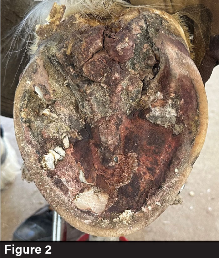
The cause of coronitis is not entirely understood (Williams 1882; Merriam-Webster Medical Dictionary; Mahon 1900; Reeks 1918). Nothing has been proven, but it has been suggested that it can be brought on by canker (Figure 2), autoimmune diseases (pemphigus foliaceus, which is a common autoimmune skin disorder in which the immune system mistakenly attacks proteins that hold skin cells together) (Knottenbelt 2011), sunlight hypersensitivity and viral diseases such as vesicular stomatitis to name a few causes (Williams 1882; Merriam-Webster Medical Dictionary; Mahon 1900; Reeks 1918; Knottenbelt 2011). One study showed that mange mite (chorioptes bovis) may be implicated (Knottenbelt 2011). Diagnosing the primary cause of coronitis is difficult, if it’s done at all (Buff & Reilly 2025).
Treatment
The underlying cause is often complex or systemic. Coronitis isn’t a primary disease, but a symptom of deeper problems.
Treating coronitis and coronitis associated with canker can be frustrating and labor-intensive for the owner. The prognosis and outlook vary because some cases have extreme forms that don’t respond to treatment, while others are milder and respond favorably.
The horse owner’s role is critical. Coronitis can worsen with wet, muddy or unsanitary conditions. They must ensure proper stall hygiene, dry footing and fly control.
Since coronitis often requires long-term, intensive care, the veterinarian and farrier are limited without active and consistent support from the owner.
Gain more insight by reading “Coronitis & Canker: Distinct but Interconnected Equine Hoof Conditions” in the July/August 2025 issue of American Farriers Journal


