Takeaways
- Coronitis is inflammation of the coronary band and is often accompanied by swelling, ulcerations, heat and pain. The hoof can flake, scale or crust.
- Chronic coronitis can predispose horses to canker, especially when combined with environmental factors.
- Coronitis and canker treatment often include trimming and balancing hooves, medications, improved hygiene and, in some cases, surgery.
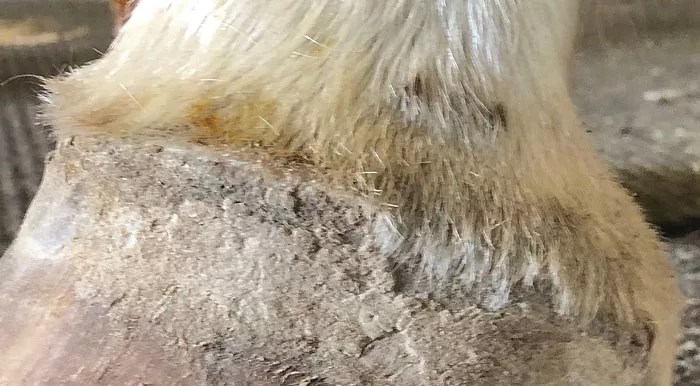
Discussions about coronitis and coronitis associated with canker have been part of veterinary literature for more than a century.
Coronitis was often described in connection with hoof infections, trauma or systemic conditions affecting the lower limb in veterinary texts from the late 19th and early 20th centuries. Practitioners observed that persistent inflammation of the coronary band could lead to chronic issues, including the development of canker, a proliferative, often foul-smelling condition characterized by abnormal tissue growth in the frog and sole.
Writers like Professor William Williams, in his The Principles and Practice of Veterinary Surgery (first published in 1882 and reprinted well into the 20th century), described hoof diseases including canker with references to preceding coronitis. They noted that poor stable hygiene, chronic moisture and neglected injuries often contributed to these problems. The link between coronitis and deeper hoof pathologies like canker was appreciated, even if the precise microbiological causes were not fully understood.
While terminology and scientific understanding have evolved, the fundamental observations are consistent: chronic coronitis can predispose horses to canker. Modern diagnostics and treatments have improved, but the challenge of preventing and managing coronitis, canker and coronitis associated with canker persists.
What is Coronitis?
Coronitis is a description of the problem — inflammation of the coronary band. (Williams 1882; Merriam-Webster Medical Dictionary; Mahon 1900; Reeks 1918; Menzies Gow et al. 2002). The term “coronitis” refers to a variety of changes in the coronary band’s appearance. The coronary band is the area at the top of the hoof where the hoof wall meets the skin. It appears as an irritation or swelling of the coronary region. At times, it can breach into the heel bulbs and frog, which appears to generate into canker (Williams 1882; Merriam-Webster Medical Dictionary; Mahon 1900; Reeks 1918; Menzies-Gow et al. 2002).
Link Between Coronitis & Canker
While early texts may not explicitly connect coronitis and canker, the progression from inflammation of the coronary band (coronitis) to more severe hoof conditions like canker was understood in practice and discussed in H. Caulton Reeks’ 1906 Diseases of the Horse’s Foot.
Contemporary veterinary literature acknowledges that chronic inflammation or injury to the coronary band can predispose horses to canker, especially when combined with environmental factors. This understanding reflects a continuity in recognizing the relationship between these conditions over the past century.
Without going in depth, canker is defined as an anaerobic (grows in the absence of oxygen) infection in the superficial epithelium of the hoof or chronic proliferative pododermatitis of yet unknown etiology (Menzies-Gow et al. 2002). The condition is often characterized by abnormal tissue growth, often with a foul odor, and is challenging to treat. It often starts in the frog and potentially spreads to other parts of the hoof (Menzies-Gow et al. 2002).
Like canker, coronitis and coronitis associated with canker does not affect the herd, only certain individual horses.
Importance of Terminology
There has been a recent tendency to confuse coronitis with Coronary Band Dystrophy (CBD). This is a misapplication of terminology. Dystrophy defines a process of hereditary progression. We don’t know whether this has a genetic lineage component (Reilly 2024; Buff and Reilly 2025).
Dystrophy defines the process of specific cells. We don’t know which cells are affected in coronitis as there are no specific histological slides of the skin or hoof for diagnosis. Biopsy results have been unrewarding and non-specific (Reilly 2024; Buff and Reilly 2025).
It is a condition of a body part/tissue that gradually weakens. We have no idea about this since there is no test on skin/hoof/frog strength lessening (Reilly 2024; Buff and Reilly 2025).
“Treatment can be frustrating and labor-intensive for the owner…”
Dystrophy can also be a nutritional or metabolic tissue issue. We have no idea whether it’s caused by some vitamins or minerals becoming too high or low. (Reilly 2024; Buff and Reilly 2025).
It is defined as any of various unrelated, noninfectious, often genetic disorders characterized by progressive deterioration or structural alteration of a body part (Beasley 2024; American Heritage Dictionary;Taber’s Medical Dictionary; Farlex 2012; Cambridge Dictionary). It’s a degenerative disorder caused by inadequate or defective nutrition (Beasley 2024; American Heritage Dictionary). There are progressive changes that may result from defective nutrition of a tissue or organ (Beasley 2024; American Heritage Dictionary; Taber’s Medical Dictionary; Farlex 2012; Cambridge Dictionary).
Signs & Causes of Coronitis
In addition to the coronary band being involved in coronitis, the ergots and chestnuts also can be involved. They can become enlarged, crusty and bleed (Williams 1882; Mahon 1900; Reeks 1918).
In a minority of advanced and long-standing coronitis cases, hoof cracks can occur. This appears to be a weakening of the hoof wall due to coronitis, but the mechanism and why this happens are unknown (Williams 1882; Mahon 1900; Reeks 1918; Reilly 2024; Buff & Reilly 2025). This was thought to be an issue with only draft or draft crosses; however, it is well-documented in many breeds.
The clinical signs include flaking/scaling, crusting, exudate or ulceration of the hoof and the coronary band area.
The coronary band swells, causing the hair to stand up or lift away from the hoof wall (Figure 1). The hair can be nearly parallel to the ground. Wart-like scaling of the periople can occur. The hoof wall loses its shiny periople appearance and becomes increasingly flaky.

Coronitis case, which is characterized by flaking and crusting of the coronary band. Esco Buff
Some horses are lame and initially mistaken for abscessing or acute laminitis. It can be painful to the touch and the horse can be lame. The coronary band is often warmer than normal (Williams 1882; Merriam-Webster Medical Dictionary; Mahon 1900; Reeks 1918; Reilly 2024; Buff & Reilly 2025). Early detection and reporting to the veterinarian and farrier are essential to prevent progression.
The cause of coronitis is not entirely understood (Williams 1882; Merriam-Webster Medical Dictionary; Mahon 1900; Reeks 1918). Nothing has been proven, but it has been suggested that it can be brought on by canker (Figure 2), autoimmune diseases (pemphigus foliaceus, which is a common autoimmune skin disorder in which the immune system mistakenly attacks proteins that hold skin cells together) (Knottenbelt 2011), sunlight hypersensitivity and viral diseases such as vesicular stomatitis to name a few causes (Williams 1882; Merriam-Webster Medical Dictionary; Mahon 1900; Reeks 1918; Knottenbelt 2011). One study showed that mange mite (chorioptes bovis) may be implicated (Knottenbelt 2011). Diagnosing the primary cause of coronitis is difficult, if it’s done at all (Buff & Reilly 2025).

A horse presents with coronitis associated with canker. Esco Buff
Treatment
The underlying cause is often complex or systemic. Coronitis isn’t a primary disease, but a symptom of deeper problems.
Treating coronitis and coronitis associated with canker can be frustrating and labor-intensive for the owner. The prognosis and outlook vary because some cases have extreme forms that don’t respond to treatment, while others are milder and respond favorably.
The horse owner’s role is critical. Coronitis can worsen with wet, muddy or unsanitary conditions. They must ensure proper stall hygiene, dry footing and fly control.
Since coronitis often requires long-term, intensive care, the veterinarian and farrier are limited without active and consistent support from the owner.
Farrier’s & Veterinarian’s Roles
Proper hoof trimming and balancing with shorter trimming cycles help reduce stress on the hoof. Remove any potential inciting causes. Have the horse owners shave all the hair about 2 inches above the coronary band as well as the feathers. Make sure they disinfect blades following the disinfecting process below.
Rasp all flaking and scaling from the hoof wall. It’s important to rasp or use a coarse sanding sponge (raspberrying) on the coronary band for topicals to work better (Figures 3 & 4) (Reilly 2024; Buff & Reilly 2025; Buff 2016). Also, trim the chestnuts and ergots for topicals to work better. Debride any canker if present.

Lightly rasp or sand the coronary band for topicals to work better. Esco Buff

A horse with coronitis pre- (left) and post-trim (right). Esco Buff
Disinfect tools after working with canker, coronitis and coronitis associated with canker. Due to the possible papilloma virus component in coronitis (proven component in canker), it’s important to know that soap, alcohol or chlorhexidine isn’t effective against papilloma virus. Tools can be flamed in the forge or treated with diluted Chlorox bleach following certain protocols (Reilly 2024; Buff & Reilly 2025; Buff 2016; EPA 2007).
An effective disinfecting procedure is to use regular 7.5% Clorox and mix 1 cup with 3 cups of water. Place the solution in a container and place the tools into the disinfection solution so they are completely covered. Soak for 5 minutes, then remove the tools, rinse them in water, dry and apply oil to prevent rusting. Make sure to wear eye protection (Reilly 2024; Buff & Reilly 2025; EPA 2007). Discard the solution after use, as it’s not potent enough after 24 hours (Reilly 2024; Buff & Reilly 2025; EPA 2007).
Remove potential inciting causes. Applying topical treatments is more effective by raspberrying the coronary band (Reilly 2024; Buff & Reilly 2025). Systemic antibiotics are often used, as necessary. The key is to treat coronitis and coronitis associated with canker like canker (Reilly 2024; Buff & Reilly 2025).
Can canker be an autoimmune disease? No blood or antibody-antigen testing has shown this — it’s only a theory (Reilly 2024; Buff & Reilly 2025).
Drugs that suppress the immune mechanisms are usually used (corticosteroids), but they have potentially harmful side effects, especially at extremely high doses. Light-dose topical retinoids and steroid creams have varying effects (Reilly 2024; Buff & Reilly 2025).
High-dose oral steroids are not recommended as they can cause laminitis and increases in papilloma virus (a canker component), bacterial counts (a canker component) or fungal counts (a possible canker component) (Reilly 2024; Buff & Reilly 2025).
Several owners that have used high-dose oral steroids on coronitis report raging canker in all feet within 2 weeks and sarcoids (papilloma component) on the ears and belly. Conclusion: This treatment is a gamble. I recommend skipping it. Non-steroidal anti-inflammatory drugs (NSAIDs) may also be prescribed (Reilly 2024; Buff & Reilly 2025).
Protocol for Coronitis
In coronitis cases, follow this protocol.
- Trim and balance the hooves.
- Have the horse owner shave all the hair about 2 inches above the coronary band, including the feathers.
- Rasp the flaking and scaling on the entire hoof wall.
- Lightly rasp or use a coarse sanding sponge (raspberrying) on the coronary band so topicals work better (Reilly 2024; Buff & Reilly 2025; Buff 2016).
- Trim the chestnuts and ergots for topicals to work better (Reilly 2024; Buff & Reilly 2025; Buff 2016).
- Deworm with one tube of Ivermectin one time a week, 4 consecutive weeks – in case of nematode (worm) aspect. Ivermectin is also an immune modulator (Reilly 2024; Buff & Reilly 2025).
- Apply topicals to the coronary bands, even those that do not appear to have coronitis. I recommend Dr. Frank Reilly’s Creative Science (formerly Equine Medical) coronitis lotion applied twice a day for 2 to 4 weeks (Reilly 2024; Buff & Reilly 2025).
- The coronitis lotion can be made with the following equipment: a blender to grind the tablets, a 4-cup mixing cup, a tablespoon, a funnel to transfer lotion to dispensing bottle, a plastic bag to hold the coronitis powder blend, a whisk and a dispensing bottle (Reilly 2024; Buff & Reilly 2025).
- Reilly made his coronitis lotion public and the recipe follows. Mix CBD powder with a gallon of basic massage lotion to make the topical. Mix 6 tbsp of aspirin powder, 6 tbsp of Doxycycline powder, grind 40 tablets of double-strength 960 Bactrim sulfa-
- trim tablets, grind 500 tablets of 500 mg metronidazole tablets, grind 200 tablets of zinc gluconate tablets and grind 100 tablets of 200 mg ketoconazole tablets. Take 10 tbsp of this powder and mix with 1 tbsp of water, 3 tbsp of DMSO liquid, and 3½ cups of the CBD and lotion mix. Mix well and apply lotion onto the coronary band twice a day for 2 weeks in a row. No wrapping needed. Let the lotion drip onto the hoof wall, frog and heel bulbs (Reilly 2024; Buff & Reilly 2025).
- The coronitis usually clears within 2-4 weeks. To prevent recurrence, utilize Reilly’s coronitis tea (Reilly 2024; Buff & Reilly 2025) applied twice a week for 30 days. Apply the tea lotion with a paint brush just below and on the coronary band. No wrapping is needed, and the horse can have normal turnout (Reilly 2024; Buff & Reilly 2025).
- Coronitis tea (made public by Reilly) (Reilly 2024; Buff & Reilly 2025) can be made at minimal cost. In a wide-mouth glass jar, mix 1 tsp of copper sulfate pentahydrate powder, 2 tbsp of 99% Dimethyl sulfide liquid, 2 tbsp of Betadine 10% povidone-iodine solution, and 2 cups of tap water. Mix until the copper sulfate crystals dissolve.
Protocol for Canker
In canker cases and no coronitis, follow this protocol.
- Debride all of the canker. I recommend learning to palpate cankerous material and flushing during removal instead of relying on visualization only.
- After debriding the canker, treat with Canker Powder and protocol (Reilly 2024; Buff & Reilly 2025; Buff 2016; Witcliffe Veterinary Pharmacy Equine Medical & Surgical Assoc.).
Protocol for Coronitis Associated with Canker
In cases of coronitis associated with canker, follow this protocol.
- Follow the protocol for canker with no coronitis.
- In 30 days, follow the protocol for coronitis only.
- In theory, both these issues can be treated at the same time; however, we have had greater success treating the canker first for 30 days, followed by treatment of the coronitis.
Treatment of equine coronitis is difficult because it often reflects a deeper systemic or autoimmune disease, affects a sensitive area and can result in permanent hoof damage and chronic pain. Long-term management and a multidisciplinary approach involving the veterinarian, farrier, other equine professionals and the horse owner are often necessary and the key to successful outcomes.
The historical recognition of coronitis and canker and their interrelation underscores the longstanding awareness of the importance of hoof health. While terminology and treatment methods evolved, the fundamental challenges of preventing and managing coronitis and coronitis associated with canker persist.
Acknowledgment
My sincere gratitude to Frank Reilly, DVM, for his valuable assistance with the 2025 International Hoof-Care Summit lecture and critical information in the development of this article.