Sooner or later, most every farrier will come across a problem foot and wonder what’s going on inside the hoof wall. So, when is it right for a farrier to request radiographs, and what’s to be expected after such a request?
Three equine veterinarians who were asked about the subject by American Farriers Journal each immediately mentioned lameness as a sufficient reason to call for radiographs. But there are many other situations that justify taking radiographs, they agree.
Veterinarian Mike Coker of Carrolton, Ga., says, “As a farrier, if I were shoeing a horse and the horse didn’t look comfortable or sound, or it didn’t move well, and there’s nothing I can see on the outside to explain this, I need to look more in depth at what’s going on within the foot. At that point, I’d want a radiograph.”
Coker, who’s also been a certifined journaeyman farrier for 17 years, adds, “If you have a hoof capsule that’s growing in an odd shape or fashion and you want to know if there is some reason internally why the capsule is growing that way, it might help to radiograph it.”
Veterinarian Mike Pownall of Rockwood, Ontario, concurs, saying that in cases of angular limb deformity leading to hoof capsule distortion, radiographs can help determine where the coffin bones are and how the farrier should trim a horse. Radiographs can help farriers determine the position of the bony column in relation to the hoof wall in other types of cases, he adds, even in some cases of long toe.
Radiographs might also be appropriate for assessing disease such as white line or conditions such as navicular syndrome, says Pownall, a certified farrier who continues to shoe horses.
He notes, “I’m working a lot with farriers in our area who are realizing the benefit of X-rays to help them with hoof balance and how they’re trimming a problem foot.”
Veterinarian Tia Nelson of Helena, Mont., says that if a lameness problem is not responding to reasonable care by the farrier or a horse is not living up to expected performance levels, it would be appropriate to request radiographs. She seldom takes radiographs unless the horse is acutely lame and doesn’t want to put its foot down, and the problem doesn’t appear to be an abscess, she says.
Be Prepared
But before you ask for radiographs, try to isolate the problem within the foot so you can offer the vet more information as a starting point.
As Coker notes, horse owners often put him on the spot by requesting radiographs without first isolating the problem. “Where should I radiograph? Where should I start? Would they like a whole body radiograph? That’s a lot of radiographs and would take a lot of time and money to do,” he says.
But even after offering information, a farrier should not expect a veterinarian to walk in and immediately start taking radiographs. “No vet should come out and do radiographs without doing a lameness exam first,” Coker says. “In the process of the lameness exam is when radiographs would be taken.
“The smart thing to do is to come out with the intention of finding where the horse is lame. We can determine that by using hoof testers and local anesthetic blocks. When we know where the horse is hurting, then we know what to radiograph,” Coker adds.
In fact, if the farrier is not already working with the veterinarian and the request for radiographs must be made to the horse owner, Coker says, “You might not put it in terms of requesting a radiograph. You’d tell the owner that the horse is lame or the hoof is growing in an odd fashion, or the horse is not comfortable or sound, so more information is needed.”
Coker suggests that a farrier talk directly with the veterinarian after clearing such discussions with the horse owner. “Sometimes when a message goes from the farrier to the owner and then to the veterinarian, or vice versa, the message can get a little confused. It would be a good idea for the farrier and vet to talk directly,” he says.
Go Ahead, Ask
When a farrier knows that a veterinarian has already examined a laminitic or foundered horse without taking radiographs, the farrier can reasonably request that the shots be taken.
“It would probably be a smart thing at that point to ask for radiographs,” Coker says. “I can understand the farrier wanting to see radiographs so he knows if he’s dealing with any rotation. I’d want to talk to the veterinarian and ask for some radiographs to be taken, or ask if there is some reason the vet doesn’t want to take radiographs.”
If the veterinarian has already taken radiographs of a problem hoof, farriers should not hesitate to ask to review the shots for helpful information, according to Pownall.
“I think that’s absolutely essential; the farriers are part of the team with the vet and the horse owner,” Pownall says. “It’s great to involve the farrier and tell him what I’ve seen, but a picture is worth a thousand words. It’s one thing to describe it, but if the farrier can see what’s going on, that’s beneficial.”
Put It In Context

WORKING TOGETHER. Veterinarian Chris France and farrier Jamey Carsel look over radiographs as they plan a treatment for a foundered horse at Pea Ridge Veterinary Clinic in Pea Ridge, Ark., in this 2003 file photo.
Although radiographs are valuable tools, farriers should not consider them the only input for determining how to care for a problem hoof.
Coker cautions: “A radiograph in and of itself doesn’t tell you very much if you’re not looking at the rest of the horse. You also have to look at the hoof capsule and how it’s growing, and what shape and condition it’s in. Look at the legs. Are they straight or are they crooked? Does the horse have any conformational problems in the limbs?
“Don’t focus on the radiographs and forget the rest of the job. Use your five senses first, before you look for radiographs. Look at and get your hands on these horses and watch them move,” he says.
One reason that hoof care cannot be based solely on radiographs is that the radiographs can be misread or misinterpreted.
Pownall cites two hoof conditions that can commonly be misinterpreted in radiographs. He says some navicular bones that look healthy in radiographs are actually degraded and causing navicular syndrome, while other navicular bones appear to be in poor condition, even as the horse shows no outward effects. The increasing use of MRIs, another form of diagnostic imaging that can often provide even clearer pictures than X-rays, is proving this point, he adds.
Laminitis is also commonly misidentified, according to Pownall, when there is capsular distortion and the radiograph is interpreted as pedal bone rotation and founder, but in fact the line of the P1, P2 and P3 are properly positioned.
Nelson says many hoof conditions can be misinterpreted in radiographs. She recalls a case, referred from another veterinarian, that was thought to be a fractured coffin bone. It turned out that the horse was sore due to an abscess, and what were thought to be fracture lines were actually vascular channels on the coffin bone.
“That’s not an unusual thing to have come to me,” she says. “That’s probably the most common one I’ve seen.”
According to Coker, who says “reading radiographs is part science and part art,” almost all hoof problems or conditions can be confused or misread while reading radiographs that are not completely definitive.
“That’s why if it’s a single hoof involved, and no problem jumps out at me in the first set of radiographs, I’ll take shots of the opposite hoof or leg,” he says.
“Presumably, that other foot is normal, so I can compare the radiographs of the problem foot with the good foot and look for anomalies.”
Take Another Look
When farriers and veterinarians work together on problem hooves, follow-up radiographs are an option for judging the progress of the feet. But how often is too often for follow-up shots?
The appropriate frequency of new radiographic images would depend on the pathology of the particular foot, the veterinarians agree, and they are open to input from farriers.
“Even if it’s laminitis and you’re trying to derotate the hoof capsule, and you want to know how it’s progressing, it would still depend on how quickly you’re trying to make the changes. Taking shots every shoeing or every other shoeing would be appropriate in that kind of case,” Coker says.
“If it’s another kind of hoof problem, then probably not quite so frequently,” he says, “because other things are not going to change so fast within the hoof. It’s done as you think is necessary, depending on the case — the pathology you found to begin with and whether you’re seeing progression or regression.”
But, Coker says, “If I was working with a farrier on a case and he said he was stuck and needed more radiographs to judge what was happening, I wouldn’t mind shooting some more, as long as it’s OK with the client. Anytime is an OK time, as long as you think it’s telling you something and it will help the horse.”
He notes that when he works with ultrasound images to check on soft tissue such as a tendon, he might take new images as often as every 30 days. “I’m looking for progression, and I know I can see it at that pace. But if it’s something bony and it’s not changing that fast, I don’t think taking radiographs that often would help much.”
Pownall says the frequency of radiographs depends on the condition of the hoof and what the farrier and vet decide. “Every case is unique. To say new shots should be taken every 6 weeks or 12 weeks would not do justice to certain situations. It’s a team decision between the farrier and the vet.”
He adds, “It might be too soon from the vet’s point of view, but if the farrier is seeing changes in the foot, it might be warranted.”
But don’t get carried away with repeated radiography. Nelson cautions that it is not necessary to routinely radiograph feet to check for progress. “I almost never radiograph hooves, unless a hoof problem in not resolving itself within a reasonable period of time,” she says.
Sharing Images
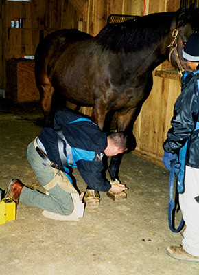
PROPER POSITIONING. Veterinarian Scott Morrison prepares a horse’s front feet for radiographs during a barn call in this 2003 file photo. Elevating the feet on wooden blocks allows the radiographs to capture the entire foot from the sole to the coronary band.
The veterinarians say they are happy to allow farriers to review the radiographs, but they note that they don’t have the option of allowing farriers or horse owners to keep the images.
As Coker says, “Legally, in my state, I have to keep those pictures for 7 years. They are part of the permanent medical record. I’m responsible for those records.”
He says farriers and clients should remember that horse owners don’t buy the actual radiographs. “They’re paying me to take the shot and read the radiograph and tell them what’s wrong with the horse. That’s what taking radiographs is about; it’s not about holding the films themselves,” he says.
Coker says the images might be sent to another veterinarian or a university for review and consultation, but the other veterinarian is obligated to return the shots.
“People outside the veterinary profession don’t have the same obligation, so vets can get into legal trouble by giving out radiographs and never getting them back,” he says.
Farriers can request copies of the radiographs to keep, but the request should be made prior to the process of creating the images.
Coker notes that he can load a second film into his radiography equipment, “so when I develop the films, I have a set for me and my permanent records and another set of shots for the owner or the farrier. I don’t mind making copies to share, as long as I know ahead of time.”
However, he will not give copies of radiographs to a farrier without the horse owner’s permission.
Coker says copies of digital radiographs can be produced easily by computer, meaning a request prior to the shooting of the images isn’t essential. However, he notes that not all veterinarians use digital equipment because it is more expensive than traditional X-ray film equipment.
Pownall also says sharing radiograph images has become easier now that the digital images can be e-mailed to farriers with notations. “There’s no excuse not to have that collaboration between the vet and the farrier going on,” he says.
Don’t Go Too Far
Although the veterinarians share images with farriers and welcome the input of the shoers, they all caution that farriers should be careful, for their own sake, not to overstep their legal bounds.
Coker warns: “It’s not the farrier’s job to interpret the radiographs, that’s the veterinarian’s job. Reading a radiograph is a practice of veterinary medicine.
“Don’t let somebody talk you into doing that. If you read a radiograph and start taking treatment actions and you’re wrong, you could be taken to court.”
Pownall agrees, noting that the law varies from state to state or province, but reading and acting on the radiograph is generally considered a medical diagnosis, which is solely the responsibility of veterinarians.
Nelson — who has been shoeing for 27 years and has been a veterinarian for 8 years — says she knows some farriers take radiographs on their own. She says it is very clear that in most states, if not all, “if you are diagnosing and treating a condition, you are practicing veterinary medicine, and you need a license to do that.”
Nelson is licensed to practice veterinary medicine in Montana, Washington and Idaho. Idaho is unusual, she notes, because an exception in the veterinary practice law allows non-veterinarians to treat lameness problems in hooved farm animals, including horses.
“I think Idaho’s practice act is very good. I’m pleased with that,” she says.
Nelson makes it clear that she favors the use of radiographs by farriers within the bounds of their state laws.
“If farriers are using radiographs to determine if the foot is in balance, I think that’s a good thing. Any tool that you can use, anything that you can have in your arsenal to try to get those feet as good as they can be is a good thing.
“You’re not at that point diagnosing, you are simply collecting data, and I don’t think that’s dangerous. I don’t think using hoof testers is dangerous. Any farrier worth his or her salt should have a good pair of hoof testers and know how to use them,” she says.
“But,” she warns, “recognize that the veterinary practice act in your state might be fraught with danger for a farrier who is interested in lameness issues in horses.”






Post a comment
Report Abusive Comment