Articles Tagged with ''MRI''

Research Journal: May/June 2016

The information, ideas and opinions expressed are those of the author and do not necessarily represent those of the United States Department of Agriculture.
Joint Injections Compared Joint injections using corticosteroids, hyaluronic acid (HA) or a combination of the two are commonly used to treat synovitis and arthritis in performance horses. In this study, researchers compared the efficacy of one type of corticosteroid (triamcinolone acetate, TA) with and without HA for the treatment of lameness, localized to one limb only that responded to a diagnostic joint block. Eighty horses of various breeds were enrolled in the study with follow-up at 3 weeks by re-examining the horses and 3 months by an owner questionnaire. No other medications were permitted during the first 3 weeks of follow-up.
Read More

Research Journal March 2016

The information, ideas and opinions expressed are those of the author and do not necessarily represent those of the United States Department of Agriculture.
Researchers in Canada used cadaver limbs to study the effects of hoof angles and loading patterns on joint surface contact areas in the fetlock. Eight limbs from Standardbred horses that died for reasons unrelated to this study were fitted into a mechanical pendulum device to simulate hoof strike at a trot. Strips of pressure-sensitive film were placed across the joint surfaces inside the fetlock to measure contact areas, and the limb was loaded while simulating flat, toe-first and heel-first landings.
Read More

Research Journal

A 10-year study in the United Kingdom investigated the relationships between clinical signs, response to nerve blocks, X-ray and magnetic resonance imaging (MRI) findings for horses diagnosed with navicular disease. The 4,618 horses examined were presented with a complaint of forelimb lameness and received a typical clinical workup prior to MRI.


Read More

Briefings

When laminitis is a major concern and a damaged hoof wall is restricting blood flow to the laminae and resulting in further inflammation, Amy Rucker says it may be time for a hoof resection.
Read More

Briefings

Because of the typical lack of balance in a low heel and long toe situation, French hoof researcher J.M. Denoix says the foot often loses the ability to restore itself back to normal. This is due to permanent damage to various internal structures such as the way the navicular bone is connected to P2 and P3.
Read More
DiagnosticImaging_Opener.jpg

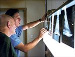
Using Diagnostic Imaging to Pinpoint Foot Problems

The vital information that technology can provide is another reason for farriers to work closely with veterinarians
Many new diagnostic techniques and imaging modalities have come into use and it can often be challenging to understand which ones might be best suited for a given lameness problem. It is also important to know when to use these tools.
Read More

Top Articles

Current Issue

View More

Current Issue

View More

Must Read Free Eguides

Download these helpful knowledge building tools

View More
Top Directory Listings