Takeaways
- Performance laminitis is characterized by inflammation, micro-damage and instability of the coffin bone within the hoof capsule. This condition is often the result of sustained mechanical stress.
- Stress has a profound impact on the foot. Prolonged hormonal stress can weaken connective tissues, impact immune response, reduce blood flow to the hoof and weaken the basement membrane — all of which can lead to laminitis.
- To reduce laminar strain, assess the root cause of the laminitic event.
Performance laminitis is characterized by inflammation, micro-damage and instability of the coffin bone within the hoof capsule. This condition is often the result of sustained mechanical stress — including coffin bone compression against the inner wall, torsional forces and repeated chemical or physical insult to the laminae.
More significant contributors include a steady cocktail of chemical exposure from misused “calming” medications such as dexamethasone, magnesium sulfate, gabapentin, tryptophan and assorted hormones; questionably necessary supplements tossed in like seasoning; frequent joint injections; and conditioning programs that sometimes begin only after the show season is already underway. The result is often a horse with gut issues, low-grade ulcers or a general sense of malaise — yet still expected to perform at their peak.
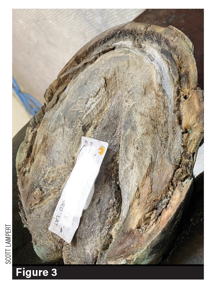
A lack of laminae strength allows the coffin bone to descend and result in a dropped sole.
The Stress Cascade
Stress has a profound and often underestimated effect on the laminae of the equine hoof. These structures — comprising both sensitive (dermal) and insensitive (epidermal) laminae — are responsible for suspending the coffin bone within the hoof capsule. When subjected to ongoing physical, metabolic, emotional or environmental stress, a cascade of physiological changes can weaken this vital interface and compromise hoof integrity (Figure 3).1
1. Hormonal response. When a horse experiences stress from pain, travel, heat, injuries or training pressure, the hypothalamic-pituitary- adrenal axis — the body’s built-in stress response system — is activated.
- Cortisol release. Chronic stress causes prolonged elevation of cortisol, the body’s primary stress hormone. In the onset, cortisol is beneficial. It helps the horse survive and manage initial stressful events or conditions. However, if the stress continues or escalates day after day, this system stays turned on too long. That’s when cortisol turns adverse and starts to weaken the body by suppressing and weakening connective tissues, dampening immune response — which increases susceptibility to inflammation, slow healing and intestinal discomfort and problems.2
2. Vascular effects. Stress- induced vasoconstriction reduces blood flow to the foot, which results in decreased oxygen and nutrients. Hypoxia (low oxygen) and ischemia (blockage of blood flow) contribute to inflammation and cellular breakdown.3
3. Inflammatory cascade. Stress also activates systemic inflammation, which gradually sensitizes and compromises the laminae. This system condition weakens the basement membrane zone — the interface where dermal and epidermal laminae interlock. It’s the biological Velcro that helps hold the coffin bone to the hoof wall. The result is mechanical instability, increased susceptibility to shearing forces and detachment of the laminar bond (Figure 4).4
Digestive compromise leading to toxemia — toxins entering the bloodstream. Stress reduces gut motility and microbiome balance, providing an environment for digestive conditions like “leaky gut syndrome,” a heightened gut permeability that allows endotoxins and bacterial byproducts to enter circulation, contributing to systemic inflammation. These circulating toxins target laminar capillaries, inducing edema (accumulation of excess fluid), inflammation and further weakening of the laminar interface.
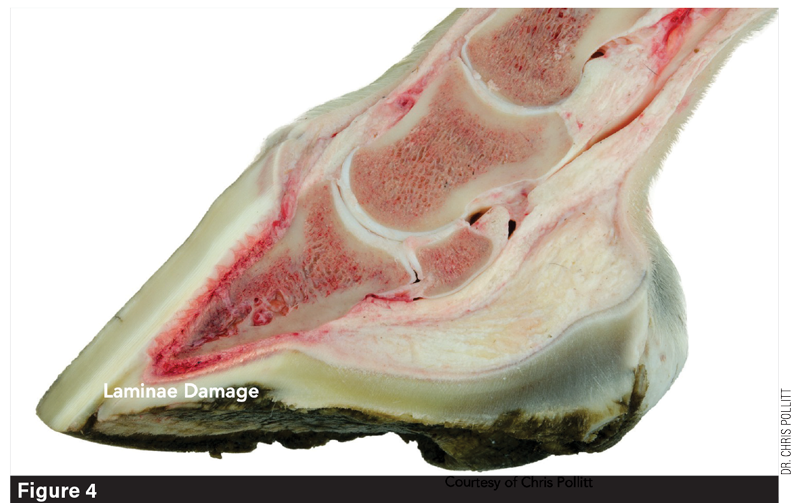
Stress-induced systemic inflammation gradually sensitizes and compromises the laminae. It weakens the basement membrane zone, leading to mechanical instability, increased susceptibility to shearing forces and detachment of the laminar bond.
Mechanical Contributors & Misconceptions
While poor trimming or “hoof balancing” is often blamed, I believe this factor is frequently overstated. In my experience, more critical contributors are the decisions made around shoe selection and fit. Overly thick or wide shoes add unnecessary weight and can fatigue an already compromised system — especially on synthetic footing, which typically consists of silica sand, fiber and wax.
A 2009 preliminary study found that while synthetic surfaces may reduce overall GRFs, their high coefficient of friction significantly shortens the slide phase. This loss of a key shock-dissipating mechanism (slide phase) leads to excessive flexion of the distal interphalangeal joint (coffin joint) and hyperextension of the metacarpal joint. As a result, tensile load increases on the laminae, collateral ligaments and supporting soft tissues — particularly the deep digital flexor tendon at its attachment on the flexor surface of the coffin bone, impar ligament, as well as the entire suspensory apparatus.
Torsional stress on these structures is further amplified during turns or circular movement, significantly increasing the risk of strain and injury. Training or disciplinary methods that rely on aggressive turning or prolonged lunging — particularly sessions lasting 20 to 60 minutes — can substantially exacerbate this risk.
Pads and packing — commonly applied to relieve perceived ground force pain or to support declining hoof quality — may offer temporary relief but often introduce pressure that exacerbates inflammation. Likewise, shoes fitted too short can create a “drop-off” effect, failing to adequately support limb leverage during the landing phase and increasing strain on the laminae.
Chemical poisoning, nutritional overload, performance stress, loss of hoof slide and repeated steps that strain the laminae over the course of the shoeing cycle all contribute to chronic micro-damage — a slow, compounding deterioration I refer to as a “death by a thousand cuts.”
Practical Steps: Reducing Laminar Strain
Treating performance laminitis requires listening to the horse, trusting a rider’s feel and being willing to honestly address mechanical and systemic causes. Key strategies include the following.
- Conduct metabolic assessments. Ensure your veterinarian performs regular metabolic evaluations, including measuring insulin and adrenocorticotropic hormone levels, to identify underlying conditions such as insulin dysregulation or Cushing’s disease. Early detection and management of these metabolic factors can significantly reduce the risk of laminitis and improve overall hoof health.
- Assess systemic stressors. Evaluate nutrition, supplements, performance medications and joint injections. Consider the unique effects anxiety inflicts by travel, nutrition, medications and disrupted rest.
- Understand GFRs. Synthetic footing typically reduces the necessary slide phase, altering proper limb position and hoof loading — creating body soreness and physical breakdown and likely introducing gait adaptations.
- Correct mechanical issues. Avoid overemphasizing lateral hoof landing or trimming based on perceived radiographic “balance” at the expense of true function. Prioritize facilitating appropriate breakover. Long, flat toes increase laminar strain — especially on hard or structured surfaces such as synthetic footing, pavement, concrete or compacted gravel, all of which are common at show facilities.
- Supportive shoeing. Use pads and pours judiciously, weighing their benefits against potential mechanical drawbacks. The ability to use hoof packing at night provides orthotic support and anti-inflammatory benefits.
- Cold therapy. Ice or cold-water therapy remains one of the most effective methods for reducing inflammation and promoting circulation. A combination of ice and water offers dual benefits — sustained cooling and mild hydrostatic pressure. While time-consuming, this approach can be highly effective in managing early laminar inflammation.
- Address systemic inflammation. Use targeted nutritional support, appropriate NSAIDs and prioritize needed rest.
- Overused or misused medications. Misusing “calming” or performance-enhancing medications and supplements often fuel the laminae and physical breakdown. Consider limiting or removing entirely.
- Alternative therapies. Low setting pulsed electromagnetic field (PEMF) or magnet therapy may enhance blood flow and reduce inflammation in subtle cases but avoid in consistently painful acute cases to prevent adverse reactions. Theraplate therapy may offer analgesic benefits and support healing, although its numbing effect warrants caution. Shockwave therapy is generally not recommended for foot-related inflammatory issues due to the risk of exacerbating inflammation within the confined hoof capsule.
References
Setterbo JJ, Garcia TC, Campbell IP, Reese JL, Morgan JM, Kim SY, Hubbard M, & Stover SM (2009). Kinetics of the forelimb in horses circling on different ground surfaces at the trot. American Journal of Veterinary Research, 70(10), 1220–1229. doi.org/10.2460/ajvr.70.10.1220
Gain more insight by reading “Understanding Laminae Strain in Sport Horses" in the September/October 2025 issue of American Farriers Journal.


