This is the first in a series on straightness and its effects on the equine hoof.

Articulation is one of those words that reverberates with multiple meanings. In this first installment of a series concerning straightness and its effects on the hoof, I intend to supply not only language by which you can articulate the relevant concepts but also to convey a multifaceted concept of all that “straightness” encompasses.

Straightness — or its opposite, crookedness — permeates all aspects of the rider’s experience, but also the farrier’s; and yet so subtly that neither the rider nor the farrier, nor even the veterinarian, may be aware that crookedness is the root cause of many apparently unrelated difficulties.

Takeaways

  • Crooked horses experience discomfort during movement, which can lead to ways to avoid work and behavioral and physical compensations.
  • It’s normal for the hind limb plane of assessment to orient outward to the front/inward to the rear because the horse must swing the hind limb forward so the stifle clears the rib cage and belly.
  • To effectively assess conformation, a farrier must determine whether there is offset, deviation, or rotation at the knee, ankle and coffin joints. Then, determine the orientation of the limb as a whole with respect to the midline plane of the body.

Over the century and a half since modern farriery and veterinary practice came into being, straightness generally has been neglected as a biomechanical fundamental, and yet straight carriage is crucial to athletic performance. No matter the discipline, a crooked horse has great difficulty performing to its full potential; experiences discomfort during movement; and therefore, will seek ways either to avoid the work it is being trained to do or else to escape the full athletic demand through various behavioral or physical compensations. The ramifications of these include, but are by no means limited to, changes in tubule orientation and hoof shape. Horses commonly compensate by alterations in gait that manifest as diminished quality of movement. Over time, compensation for crookedness usually eventuates in lameness.

Anatomical-Terminology

Click to enlarge

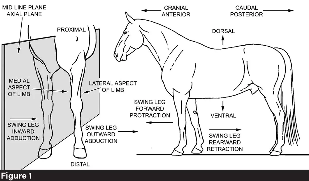
Anatomical terminology that will be referenced throughout this article. Photo by: Dr. Deb Bennett

Alignment at the Joints

Straightness is complex, so to gain a comprehensive understanding, it is helpful to take one aspect at a time. The first aspect to consider is (pardon the pun) very straightforward: the articulation of the joints of the limbs. Joints can either be “straight” — that is correctly articulated — or else “crooked,” meaning that the bones above and below a given limb joint are misaligned. Throughout this discussion, please refer to Figure 1, which explains anatomical terminology.

misalignment

Click to enlarge

The three ways in which a joint can be misaligned: (A) deviation; (B) offset; and (C) rotation. Photo by: Dr. Deb Bennett

There are a dozen picturesque words in the horsemanship vocabulary that describe limb misalignments: bench-kneed, splayfooted, knock-kneed, curby, calf-kneed, sprung, cow-hocked, pigeon-toed, upright pasterns, coon-footed, flat-footed, club-footed, windswept and bowlegged. Despite this variety, there are only three ways in which a joint can be misaligned: the bones above and below can be offset, rotated, or deviated as Figure 2 shows. Any distal limb joint can manifest one, two, or all three types of misalignment at once. Note that the diagram can represent any joint, so calf knees, cow hocks and knock-knees/bowlegs (the veterinarian’s valgus/varus) all fall into the category of deviations, whereas splayfoot and pigeon-toe are the product of rotation plus deviation at the ankle joint, and both bench knees and curby hocks represent offsets.1

Ulna-Dark-Tone

Click to enlarge

In horses, the ulna (dark tone) is completely fused to the radius. The lower end of the ulna forms the lateral portion of the distal end of the radius-ulna composite bone. This illustration also shows the deep C-shape of the glenoid cavity, the hollow in which the distal end of the humerus rotates. The articulation is tight and movement capability is constrained to hinge-like opening and closing, making elbow joint dysfunction rare. Photo by: Dr. Deb Bennett

Supination and Pronation

Misalignment of the knee, hock and ankle joints is the major focus of traditional conformation study. Unfortunately, this approach ignores the structure and function of all the bones and joints of the horse’s body above the level of the gaskin and forearm — yet the movement possibilities inherent in the design of the shoulder, stifle and hip joints govern function in the distal limbs. They therefore also govern hoof strike and breakover, which are the major factors that influence hoof form.

Femur-Head

Click to enlarge

The articulation of the head of the femur with the hip socket (acetabulum) is a good example of a ball-and-socket design. Blue arrows show how the femoral head fits into the socket; the red line is the top edge of the plane of assessment passing vertically through the femoral shaft. Note that its orientation to the pelvis is oblique so that the lower end of the femur and the stifle joint orient outward to the front. The view is left lateral, with the horse’s head to the left. Photo by: Dr. Deb Bennett

Realizing that the shoulder joint has something to do with the conformation of the forelimb — and thus the stance and movement of the horse — compels us to consider how and why. In the limbs of horses, the joints at the shoulder and hip are the only ones that can swivel more than a few degrees. Because horses are large and rather heavy animals, they need joints that will not twist or wobble, especially during fast locomotion.

Stifle-Fig-5

Click to enlarge

The stifle has the most complicated design of all the joints in the horse’s body and is, therefore, the least stable joint requiring the most “strappage” (see Figure 6). The joint is sharply flexed in this frontal view, which shows the end of the femur above and the upper end of the tibial shaft below. The patella, which would normally sit at the front of the joint in the deep groove between the medial and lateral ridges of the distal femur, has been removed to make the main portion of the stifle articulation visible. Note how the tibial spine extends upward into the intercondyloid fossa. Blue arrows show that the plane of articulation is somewhat tilted. The red line is the forward edge of the plane of assessment as it passes through the shafts of the tibia and femur. Photo by: Dr. Deb Bennett

Articular-Surface-fig-6

Click to enlarge

This view looks straight down upon the articular surface of the proximal end of the tibia to show the many ways in which the stifle articulation requires stabilization. The menisci (blue) are fibrocartilagenous pads, which cushion the femoral condyles but also serve to “seat” them in cups. The cruciate ligaments between the menisci are extremely tough and serve to hold the bones together while also permitting a slight amount of rotation at the stifle joint. There are strong collateral ligaments (shown) and external to these, the tendons of several large muscles (femoral biceps, the hamstrings, the gastrocnemius) (not shown). The joint is further stabilized by the quadriceps tendon, the patella and the patellar ligaments (also not shown). Photo by: Dr. Deb Bennett

A good way to see the biomechanical problem horses are up against is to think of strapping a set of short stilts — about the length of cannon bones — to your feet. How tightly would they have to be strapped on before you would feel safe running a sprint while wearing them? In horses, every joint below the shoulder and hip socket is structured to open (extend) and close (flex) with as little wobble as possible. To achieve this, they have ridge-and-groove construction (ankles, hocks), tightly contoured articulation (elbow joints), stack-of-blocks architecture (knees), or internal cruciate ligaments (stifle joints) — and all of them are tightly bound and stabilized by short, collateral and annular ligaments as well as overpassing tendons (Figures 3-7).

Hock-Joint-Fig-7

Click to enlarge

The hock joint is much more stable than the stifle thanks to the tongue-and-groove design. It is formed by two large bones, the astragalus (tibial tarsal) and calcaneum (fibular tarsal), plus a group of small quadrilateral and pancake-like bones that fit together tightly and are bound together by short, extremely tough ligaments. These bones are below and behind the astragalus and calcaneum and serve merely to make the hind limb longer; they are not designed for movement. The high trochlear ridges of the tibial tarsal fit into deep grooves in the distal end of the tibia to form the mobile joint of the hock, where flexion and extension occur. Blue arrows highlight the oblique orientation of the joint, while the purple arrow shows how the calcaneal shaft diverges from this. The red line through the tibial tarsal and shaft of the hind cannon bone is the forward edge of the plane of assessment, while the red line through the distal end of the tibia is the lower edge of the plane of assessment as it bisects the shaft of the tibia. Photo by: Dr. Deb Bennett

Rotation in the lower part of the equine limb is not only inhibited by the design of the joints, but made completely impossible by the architecture of the bones themselves. The forearm in humans and most other mammals is structured by two separate bones, the radius and ulna (Figure 8). Humans, monkeys, and raccoons can hold objects between the hands. This involves making a gesture called supination, in which the palm of the hand is turned inward or even upward. Supination is made possible by the cylindrical head of the radius rolling upon a notch in the shaft of the ulna. When the palm is pronated (turned downward), the forearm bones are crossed; when it is supinated, they uncross at the lower end (Figure 8).

Pronation-supination-fig-8

Click to enlarge

Pronation and supination of the forelimb are possible in humans because the radius and ulna bones are separate. In the act of pronation, the cylindrical proximal end of the radius swivels against a notch in the ulna located just below the distal end of the humerus. Photo by: Dr. Deb Bennett

In horses, however, the ulna is fused to the radius in the crossed or pronated position — which prevents supination (Figure 3). Fusion of radius and ulna is found in most hoofed species that escape predators by running, i.e. not only horses but cows, goats, giraffes, zebras, moose, elk, yaks and wildebeest. And it’s a good thing, too. Consider what would happen to the tendons of the flexor musculature if the cannon bones and fore hooves of a galloping horse suddenly swiveled around (supinated) so that the heel bulbs faced medially or even forward.

While fusion of the radius and ulna averts disaster during fast movement, it also means that no joint of the horse’s forelimb other than the shoulder joint has the capability for rotation or “swiveling.”

In the hind limb, the situation is a little different because the fibula is much smaller than the tibia and plays no part either in limb support or movement. Rotational movement at the level of the hock is instead mainly prevented by the deep tongue-and-groove articulation between the distal tibia and the tibial tarsal (astragalus) bone (Figure 7). A small amount of rotational movement is permitted at the stifle; the X-configuration of the cruciate ligaments permits the spine of the tibia to rotate in a socket in the distal end of the femur (Figures 5 and 6).2 However, the major rotational movement of the hind limb is confined to the hip (coxofemoral) joint. Both the shoulder and hip joints have ball and socket architecture that readily permits rotation (Figures 4 and 9).

Toeing-Fig-9

Click to enlarge

Three views of the left shoulder joint (between the scapula above and the head of the humerus below) as seen from directly above, with the horse’s head to the top. The dome-shaped head of the humerus is large compared with the glenoid cavity in the distal end of the scapula into which it fits. In life, this disparity is made up for by a fibrocartilagenous “gasket” (not shown) that flares out from the rim of the scapular glenoid to cover most of the humeral head. In this illustration, the scapula remains in the same position while the humerus swivels outward (A), occupies a neutral position (B), or swivels inward (C). Rotation at the shoulder joint is the main cause for toed-in or toed-out stance. Photo by: Dr. Deb Bennett

Limbs as Three-Dimensional Objects

Before we consider the down-limb effects of rotation at the shoulder and hip joints, it’s necessary first to learn to look at limbs as they really are. Anything that has height, width and depth is a three-dimensional object, and that includes the horse’s limbs (Figures 10 and 11). This becomes obvious when we remove the forelimb from a carcass — and here, I mean the entire forelimb from the shoulder down. This is done by cutting through all the muscles that connect the scapula and humerus to the neck and ribcage. The limb then can be lifted and set aside on the dissecting table, where its thickness can be measured with calipers. The maximum thickness of the equine forelimb is found at the level of the triceps muscle above the elbow, and in a full-sized horse of 15 to 16 hands will measure about 8 inches.

Plane-of-Assessment

Click to enlarge

The plane of assessment in the forelimb: (A) toed-out stance; (B) toed-in stance. Since in these horses the plane of assessment cleanly bisects all the bones below the elbow — there is no significant offset, rotation, or deviation at any joint — both animals have perfect articulation and perfect structure. Toeing-in and toeing-out represent problems of stance, not structure. This article seeks to help you learn to differentiate these two factors, both of which contribute to forelimb conformation and function. Photo by: Dr. Deb Bennett

The hind limb is more difficult to remove, partly because the investing musculature is much thicker. The major difficulty, however, is in removing the femur from the hip socket (there is no socketed connection between any part of the forelimb and the thorax3).

The head of the femur is held into the hip socket by a short, round and very strong ligament; a knife with a long, thin, flexible, hooked blade is needed to cut this cleanly. There are also ligaments and strong tendons connecting the femoral neck and shaft to the rim of the acetabulum. Once the hind limb has been cut free, its greatest thickness is at the level of the middle of the thigh. A measurement taken there encompasses not only the powerful biceps muscle complex and the part of the quadriceps that lies on the lateral side of the femur, but also the thick adductor muscles on the medial side. Thickness of the hind limb varies more than does thickness through the forelimb but is typically at least 15 inches in a full-sized horse and can be as much as 25 inches.

Hind-Limb-Fig-11

Click to enlarge

The plane of assessment in the hind limb. Note that the horse with perfect articulation, in which the plane of assessment cleanly bisects all the bones and joints from the hip socket down, is view D, not view B. Note the difference between the cow-hocked horse in view A and the correctly built horse in view D. Photo by: Dr. Deb Bennett

Measuring the thickness of carcass limbs vividly highlights the fact that the individual limbs are three-dimensional objects, which in life cannot only flex, extend and swing but can also turn inward or outward.

Veterinary textbooks, manuals published by breed associations and riding clubs and hundreds of books and magazine articles on conformation have repeated an error that first appeared in an otherwise excellent work, “The Exterior of the Horse” written in 1890 by French veterinarians Armand Gouboux and Gustave Barrier. The error is embodied in two famous drawings — still repeated everywhere — which first appeared in their book (and reproduced here as Figure 12). The drawings imply that limb conformation can usefully be assessed by visually dropping a line through the limb. Unfortunately, this approach is not effective because a real horse limb is not a two-dimensional engineer’s drawing, but instead, as we have seen, it is a three-dimensional object that has thickness, as well as height and depth. As such, the proper way to find out the alignment at the joints, as well as the orientation of the limb in space, is to bisect the limb not with a line, but with a plane (Figures 10 and 11).

Toeing-In and Toeing-Out

The orientation of a given limb in space defines its orientation with respect to the midline plane of the body (Figure 1). In thinking about the implications of this, let’s focus first on the forelimb, because insofar as the forelimb and hind limb have different general functions — the hind limb to generate thrust and the forelimb to receive the bodyweight — outward or inward rotation of the limb as a whole will have different functional effects in the forelimb than in the hind limb.

Fig-12-Barrier-Drawings

Click to enlarge

Gouboux and Barrier’s drawings were first published in 1890. The concept they convey is bogus; no normal horse stands with its hind hoofs facing straight forward. Note the subtle twist in both the hock and hind ankle joints, the same twist that is created by any attempt to force a normally conformed horse (Figure 11, view D) to stand this way, i.e. by trimming one wall of the hoof lower or by the application of shoes with trailers. Some horses do naturally stand with the fore hooves facing straight forward, but this is not ideal. Because the diameter of the rib cage rapidly increases from the point of the breast back to the level of the girth, and the shoulder blade and arm must travel along this surface whenever the forelimb is drawn back, ideally the fore hoofs would orient slightly inward. Attempts to force a horse to toe straight forward have the same effect on the forelimb as in the hind: they twist and strain every joint. Problems of stance must be solved by addressing muscles at the level of the chest and groin and above. Photo by: Dr. Deb Bennett

Because the horse cannot supinate the forelimb, the orientation of the toe of the hoof is locked to the orientation of the elbow. In other words, if we rotate the head of the left humerus in the shoulder joint to the left (counterclockwise as seen from above in Figure 9), every part of the forelimb below the shoulder joint that faces forward rotates outward and every part that faces caudally rotates inward. Thus, when we rotate the forelimb outward, movement that originates at the shoulder joint causes the horse to “toe” outward while at the same time, and to the same degree, the elbow is brought closer to the chest (Figure 10).

The plane that bisects a horse’s limb can be twisted or broken, and you have already learned this by studying the first section of this article. A twist or break in the plane of assessment of the forelimb is nothing more than an offset, deviation, or rotation at either the knee, fetlock, or coffin joint. But if there are no twists or breaks in the plane of assessment, all the joints of the horse’s distal limb are perfectly articulated — in other words, the limb is correctly conformed and there is thus no need for “corrective trimming” or any other type of correction. Nonetheless, a horse with 100% correct limb structure may toe-out or toe-in. Why? If the joints are perfectly articulated and the hoof capsule is in normal relationship to the coffin bone,4 there is only one possible cause and that is that the forelimb as a whole has rotated either outward or inward at the shoulder joint.

Straightness-of-Limb-Fig-13

Click to enlarge

Good vs. poor judging technique. On the left is what is commonly seen at horse shows. The judge cannot possibly gain a correct idea of the straightness of the limb joints by the oblique view that standing in this position provides. On the right is the correct technique: each limb is to be evaluated separately, with the judge standing in the plane of assessment so that they can look “square” at the face of the knee in front and the hock behind. If the correct technique is important for horse show judges, it is even more important for the farrier because it’s the only way to get accurate information from the body of the horse. Photo by: Dr. Deb Bennett

The plane of assessment method is equally enlightening when applied to the hind limbs. In conformation study, it clarifies the difference between “cow hocks” and normal hind limb orientation, which in all mammals is as shown in Figure 11, view D. It is a measure of the prevailing confusion within the horse industry that correct hind limb stance is often misidentified as “cow hocks” and that horses with normal hind limb stance are penalized by supposedly knowledgeable judges at shows, while bowlegged individuals (Figure 11, view B) are often given the top prize.

Why is it normal for the hind limb plane of assessment to orient outward to the front/inward to the rear? Because when the horse moves, it must swing the hind limb forward. When it does this, the stifle must clear the ribcage and belly. In narrow-bodied horses, the orientation is at less of an angle to the midline plane of the body and in wide-bodied horses, especially those with short backs, it is necessarily greater; but the horse that habitually stands with the hind limb planes oriented parallel to the midline plane of the body — built like a workbench or a beef steer as in Figure 11, view B — is not correctly conformed and will not find such athletic endeavors as sliding stops, rollbacks, pirouettes, or high-level dressage either easy or comfortable.5

Straightness from the Top Down

This article teaches that to effectively assess the conformation and thus the athletic potential of either the forelimb or the hind limb, the farrier must follow a two-step process: first to determine whether there is offset, deviation, or rotation at the knee, ankle and coffin joints. This is done by examining first the left limb and then the right, with the judge standing in the plane of assessment as shown in Figure 13. The second step is to determine the orientation of the limb as a whole with respect to the midline plane of the body. Structure and stance are two different factors that both contribute to the horse’s apparent conformation.

Every farrier sees plenty of horses that toe out. A major causative factor is compensation, spoken of at the beginning of this article: the horse may shift effort that ought to be made by the hind limbs to the forelimbs. When it does this, it will brace the pectoral muscles — in other words, it will contract them continuously. The pectoral muscles, which pull the elbows in, are much stronger than the several small muscles on the lateral side of the shoulder joint and arm that can potentially pull the elbows away from the body wall.

Horses that brace the pectoral muscles universally toe out. Conversely, a toed-out horse that is taught to move in self-carriage and thus not overweight and misuse the forelimbs, will stop bracing the pectorals and its rider, as well as his farrier, will notice that it no longer toes out to a significant degree. I report this because as a trainer and rider, as well as a coach, I have caused it in numerous horses and so have my students. It is important because it demonstrates that changing the horse’s balance in movement and thus the way it uses its muscles quickly results in changes in its stance.

Learn More Online

Gain more insight from Dr. Deb Bennett by reading:

There are also a surprising number of horses that have an asymmetrical stance — they toe out more with one forelimb than the other. This is possible because, just as the shoulder joint and hip socket govern stance and movement in the distal limb, their functioning is itself governed by structures positioned still higher up in the horse’s body, i.e. the vertebral chain and ribcage.

The farrier who raises their eyes above the level of the ankle — and even above the level of the elbow — achieves a new level of articulation, for the spine and ribcage have far more joints than all four limbs put together. In the aggregate, they weigh more too, and to add even more complexity, they are directly impacted by the rider’s weight and choice of techniques and by saddle fit.

When we rise to the level of the spine, we are talking about “straightness” not merely in terms of correct articulation at joints but an overall bodily straightness that is termed correct carriage. How this exerts a major influence on athletic functioning — and hoof function and form — is the subject of our next installment.


Footnotes

  1. Good study photos of all these limb misalignments and conformation faults, along with much other useful information, can be found in my three-volume work titled, “Horse Conformation: Principles of Form and Function,” published in 2022. I also recommend purchasing a horse anatomy book. My top three picks for beautiful illustrations and overall usefulness are a 1950s or 1960s edition of Sisson & Grossmann’s “Anatomy of the Domestic Animals”; Goody’s “Horse Anatomy”, 2nd edition; and Kainer and McCracken’s “Horse Anatomy Coloring Book.”
  2. The spiral construction of the equine hind limb was considered in the July/August 2021 issue of American Farriers Journal under the title “Improve Your Eye for Equine Anatomy,” and rotation at the stifle joint is explained and illustrated in Vol. 3 of “Horse Conformation: Principles of Form and Function.”
  3. The “unsocketed” connection between the equine shoulder and ribcage is explained and illustrated in the January/February 2020 issue of AFJ, “Examining the Shoulder to Thorax Junction.”
  4. Abnormal relationship of the hoof capsule to the bones of the limb occurs only when the interlaminar connection has been deranged in some way. This is explained and illustrated in Volume 3 of “Horse Conformation: Principles of Form and Function.”
  5. Horses with incorrect hind limb conformation — either cow hocks or bow legs — find high-level athletic performance difficult because in every discipline it requires the horse to adopt the posture of collection. This originates with coiling of the loins, which in turn permits the horse to deeply flex the joints of the hind limb. A complete review of the anatomy and biomechanics of collection is presented in Volume 1 of “Horse Conformation: Principles of Form and Function.”