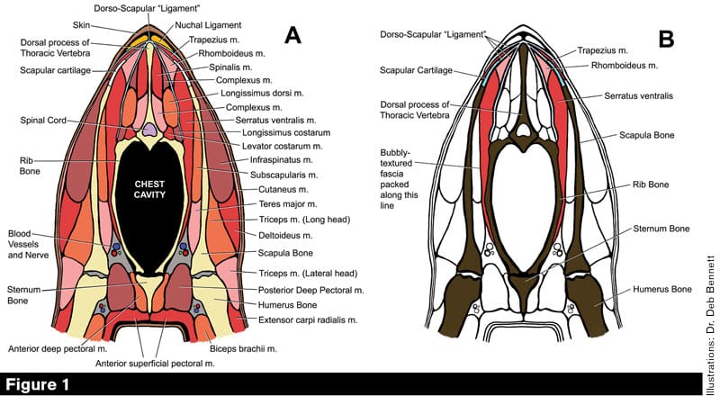
Cross sections of the equine thorax cut through the body at the level of the withers. The bones and muscles are labeled. In View A, muscles are in various shades of red and orange, loose connective tissue is gray, bones are yellow, nuchal ligament is gold, scapular cartilages are green and the DSL is in blue. View B is a simplification, with bones in dark brown for emphasis, and only those muscles that attach to the DSL are colored. Note how the forelimbs hang off the DSL. The plane where “bubble wrap” fascia is found is marked.
A cross-section cut through the horse’s thorax reveals that the equine rib cage is far from being a round barrel (Figure 1). Instead, it is shaped like a peach leaf: pointed at the bottom, with rather flattened sides. Slapped up against the flattened surface formed by the rib cage is the scapula, and the junction so formed is the connection between the horse’s forelimb as a whole and its body.
Farrier Takeaways
- The shoulder to thorax junction constitutes the upper end of the forelimb reciprocating system.
- Horses lack collar bones and there is no socketed joint between the rib cage and forelimb.
- The dorso-scapular ligament (DSL) anchors the top of the forelimb, while the serratus ventralis and pectoral muscles form the thoracic sling. Together they substitute for a socketed joint.
I say “junction” rather than “joint” because we normally think of joints as relating bones to each other by means of some kind of socketed connection. The relationship of the shoulder blade to the rib cage, however, is a muscle-bone-tendon sandwich that involves no socketed connection at all. Further, the junction is huge — in a 15-hand horse, its total area on each side measures fully 2 square feet in area. There is no other joint in the horse’s body that approaches this.
The periosteum and septa were the focus of the previous article in this series. Adding to the uniqueness of the shoulder to thorax junction is a fascinating (and seldom illustrated or discussed) septal structure called the dorso-scapular ligament (DSL). It is a “book” in which its leaves are big flaps of periosteum, and serves to anchor the forelimb to the withers.
The shoulder to thorax junction constitutes the upper end of the forelimb reciprocating system, which is our main focus in this series of articles. Many farriers hear of horses with “sore shoulders,” so in this article I’m going to take you through a virtual dissection in quest of finding out why.
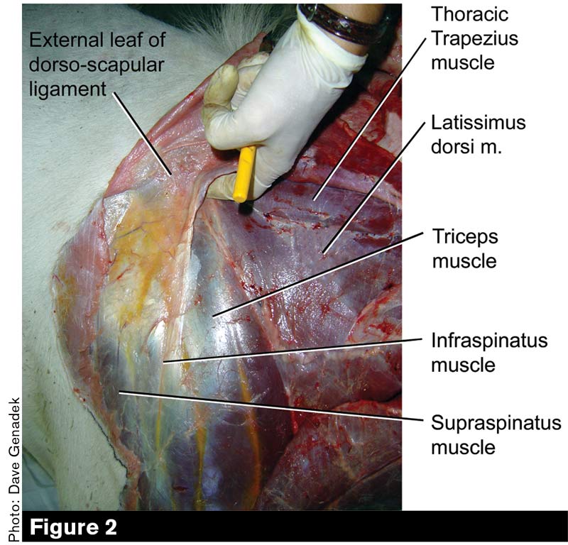
In this dissection, the animal’s neck and head are to the left. A finger is inserted beneath the most external leaf of the DSL just beneath the rear curve of the scapular cartilage. All marked muscles except the latissimus dorsi belong to the limb intrinsic functional category (see Table 2).
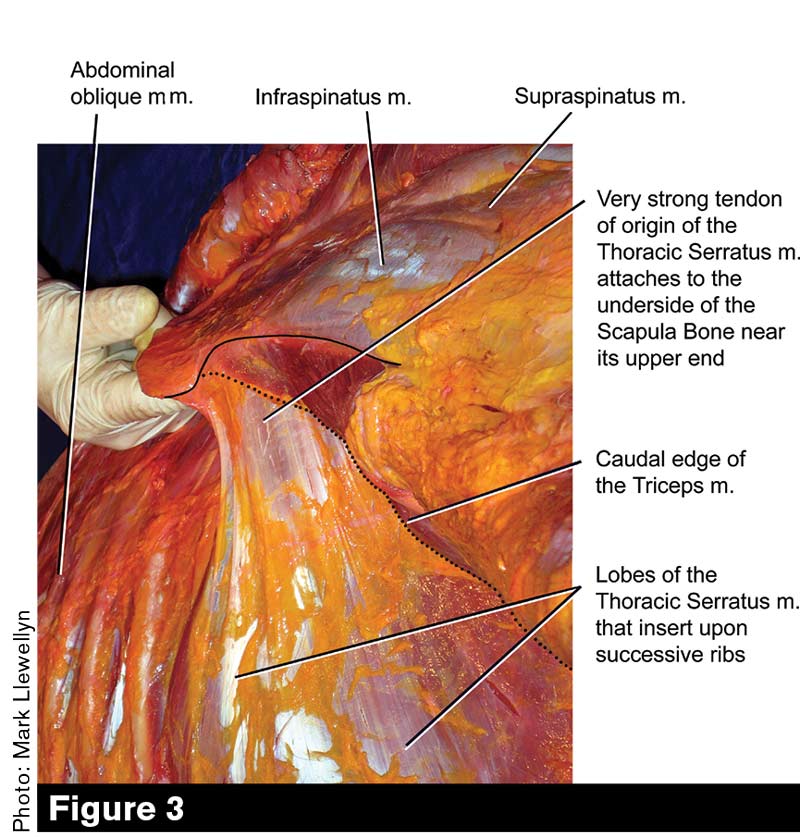
In this dissection, the animal’s neck extends 45 degrees upward to the right, and the camera looks at an angle from front to back across the curve of the ribcage. The gloved finger lifts the scapular cartilage. The rear edge of the scapular cartilage and shoulder blade proper have been highlighted by a thin black line; a dotted line defines the rear edge of the triceps muscle. The extremely large and strong tendon of insertion of the thoracic division of the serratus muscle is the main feature to appreciate.
Dorso-Scagpular “Ligament”
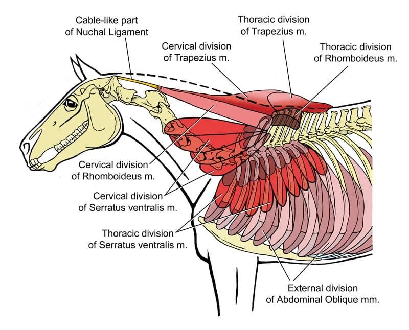
Not a ligament at all (except in the crude sense that it acts to “connect muscle to bone,” a concept we have criticized in preceding installments), the DSL is actually formed from bands of periosteum belonging to the second through the eighth or ninth thoracic vertebrae. The various bands coalesce to form a “book” of strong, thin, glistening white sheets that extend downward from the withers. Four main sheets are recognized in standard anatomy texts; three are anterior and relevant to our study of the equine forelimb. The fourth sheet is more caudal and is not discussed here or shown in the figures. Study Figures 1 to 3 to gain a good idea of the appearance and spatial relationships of the DSL and muscles that are attached to it.
The “leaves” of the DSL lie between muscle layers and function like intermuscular septa. They present a large amount of surface area, which affords muscles a place to attach. The trapezius, rhomboideus, spinalis, complexus and serratus ventralis muscles attach to one or more DSL leaves. As Figure 1B makes clear, the scapular cartilage is also attached to a leaf, and in fact the whole of the forelimb hangs from the DSL.
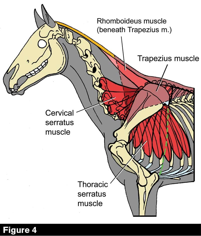
The trapezius muscle consists of two parts: a cervical part that covers the lower triangle of the neck and a thoracic part that covers the hollow of the withers. The two parts meet at a fibrous seam (“raphe”) that runs down the scapular spine. The rhomboideus muscle also consists of cervical and thoracic parts, but unlike the two parts of the trapezius, they do not mirror each other in shape. The cervical part is long and strap-like and the thoracic part (not shown here) is short and triangular. The caudal aspect of the triceps muscle, which overlies the thoracic serratus, is marked by a dashed green line.
No Collarbone Means Mobile Shoulders
The equine shoulder blade is very loose and mobile compared to that of the human, because in the horse there is no collarbone (clavicle). In humans, the pair of clavicles can be thought of as serving two purposes — picture them as spanning the shoulders out from the body, or, alternatively, you can picture them as bracing the sternum, forcing it to remain centered between the shoulders (Figures 5 and 6).
In the horse, there is nothing — not even a ligament such as found in cats and dogs — spanning between the sternum and the shoulder joints, no rigid structure of any kind, to prevent the sternum from swinging or migrating off-center; either when the horse is standing still or in motion. This fact has enormous significance to equine locomotion, veterinary lameness evaluation, riding and training technique, hoof growth and wear, and farrier care — a subject I hope to be able to go into in full detail in a future series. Meanwhile, suffice it to say that motion analyses have shown that the equine shoulder normally moves in five directions at every gait and speed: it tilts, and it slides up, down, forward and backward. The scapula scrubs against the rib cage, making motions like a sponge washing the driver-side window of your car.
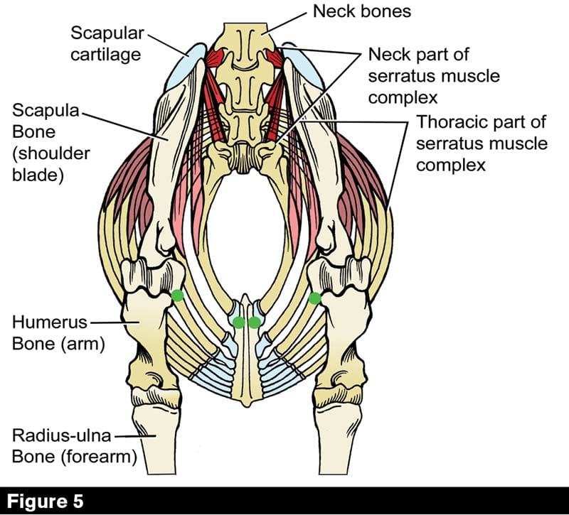
Front view of the horse’s thorax showing attachments of the serratus muscle complex. The horse lacks collar bones; in other mammals, they strut between the anterior end of the sternum and the inner aspects of the shoulder joints. To make this relationship clear, draw straight lines between the green dots.
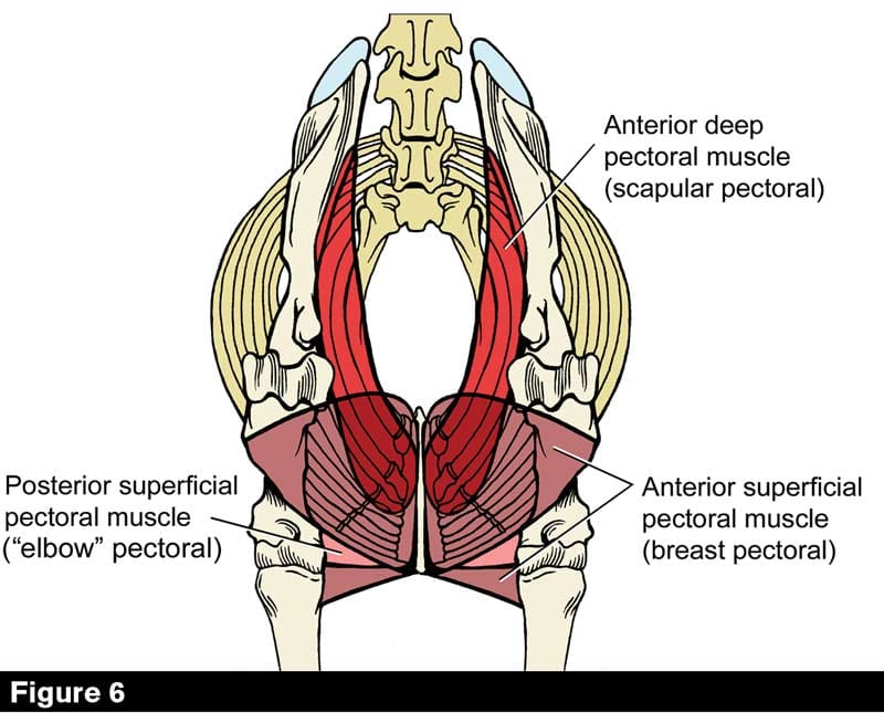
The architecture of the pectoral complex in the horse consists of four distinct divisions, three of which can be seen in this anterior view. The fourth division, the posterior deep pectoral muscle, lies behind the “elbow” and “breast” pectorals and is the muscle mass over which the girth is tightened.
Bridging Muscles
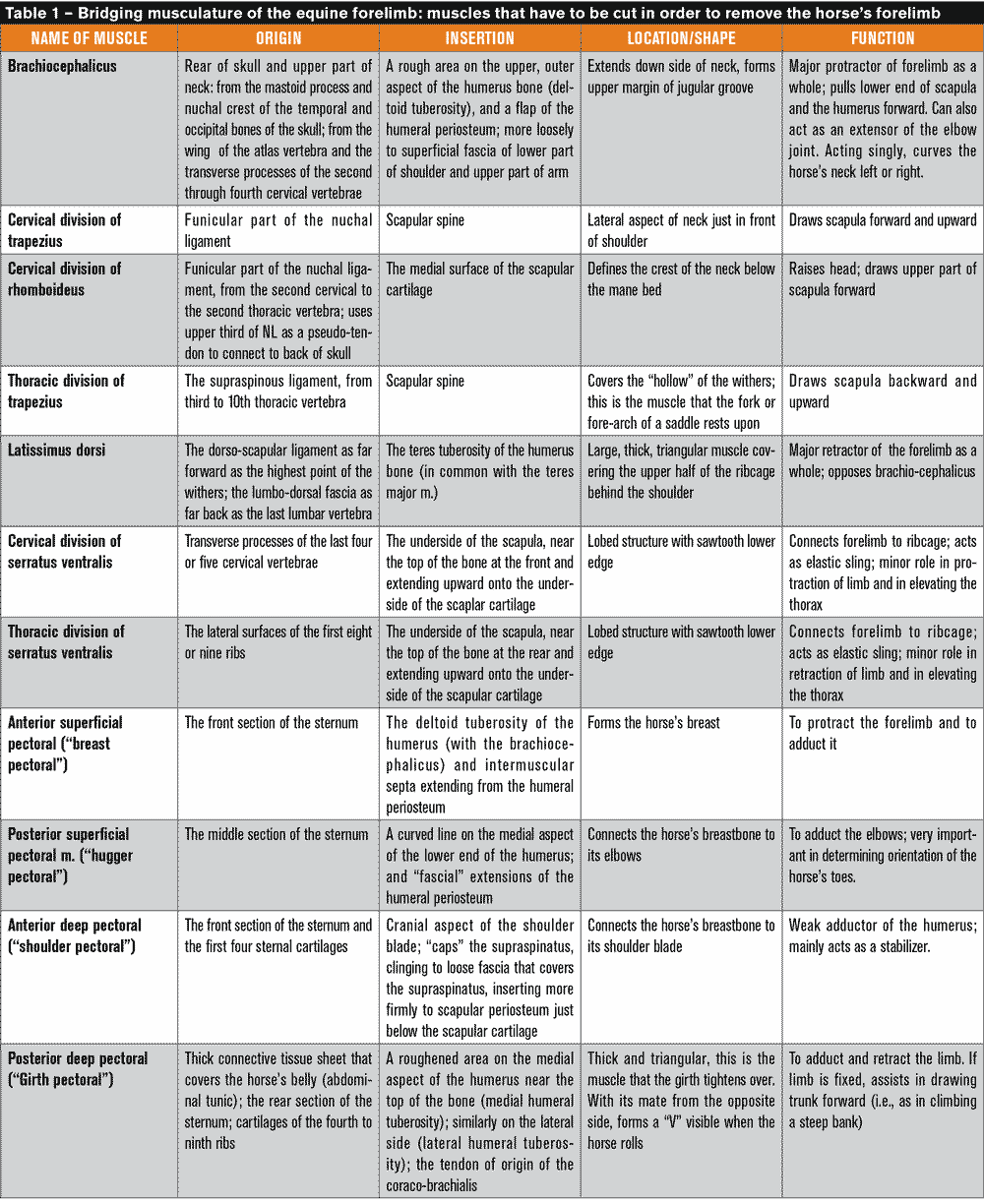
From this description, it is clear that a socketed joint would not work well to join the shoulder to the body. It would be difficult to design a joint that could provide sufficient flexibility, whereas the DSL provides both flexibility and a firm attachment. Important though it is, however, the DSL is not the only tissue that works to hold the forelimb onto the body, as there are also muscles to consider (Table 1).
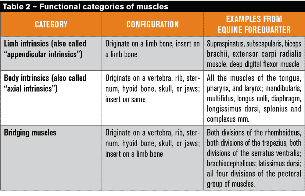
I have organizied three functional classes of muscles (Table 2): limb (appendicular) intrinsics, body (axial) intrinsics and bridging muscles. This classification is my invention, so you won’t see it referenced elsewhere. However, it can make learning the position and function of muscles much easier.
A limb intrinsic is a muscle that originates from a limb bone and inserts upon one. For example, the supraspinatus, biceps brachii or extensor carpi radialis muscles. In general, limb intrinsics are oriented up and down, paralleling the limb. By contrast, a body intrinsic is a muscle that originates from some part of the axial body and inserts upon another part of the axial body, i.e. going from a vertebra to a rib, or from the neck to the skull, jaws, or hyoid apparatus. In general, axial intrinsics are oriented fore-aft, parallel to the horse’s vertebral chain. Examples would include the longissimus dorsi, complexus or longus colli muscles.
Note that any given muscle can only affect or move the skeletal parts to which it is attached. Thus, limb intrinsics flex, extend, adduct, abduct or rotate only limbs or parts of limbs, but do not move the neck, thorax, tail, skull or jaws.
Likewise, body intrinsics flex, extend, brace, rotate or laterally flex the rib cage, neck, tail, pharynx and skull; move the tongue, effect swallowing and close or open the jaws. However, they do not move any segment of any limb.
Muscle action is normally both separate and precise — a good analogy would be the keys of a piano. Individually, each key plays only its own note. To make music rather than mere noise, each note must be related in some meaningful way to others. The pianist must coordinate the movements of her fingers to play each key at the right time. Likewise, cogent, smooth, purposeful, and athletic movement requires a coordination of muscle contractions, which is normally provided by the nervous system.
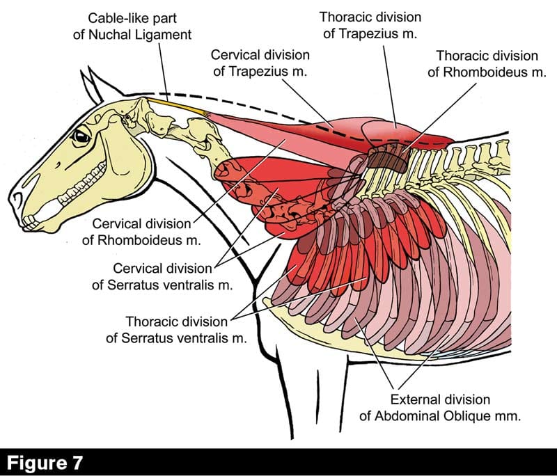
The third functional category of muscles is termed “bridging” because these muscles bridge from the body to the limbs. They are the muscles that move the limb as a whole — for example, drawing the entire forelimb forward or backward as opposed to merely folding or unfolding the carpal or shoulder joint (that would be the job of limb intrinsics). Bridging muscles are also the tissues that connect the limbs to the body. If, in a wet lab, we wish to remove the horse’s forelimb to see the structures that lie between the shoulder blade and the rib cage, we will have to cut through all the muscles which bridge from the thorax to the forelimb (Figures 7 and 8).
While all the bridging muscles function to hold the forelimb onto the body, as well as to move it, the most important of these is the serratus ventralis muscle (Figures 9 and 10, also see Figures 5 and 6). If all the other bridging muscles were cut, the forelimb would still be firmly attached to the body by means of the extraordinarily strong tendons of insertion of this muscle.
Study this figure to understand the structures in Figure 8. The cut ends of the cervical and thoracic rhomboideus muscles and cervical and thoracic serratus ventralis muscles lie under the shoulder blade in the living animal. Pectoral muscles are not shown in this view. The skeleton has been drawn directly from a photograph of a mount in my laboratory, the same animal under dissection in Figure 8.
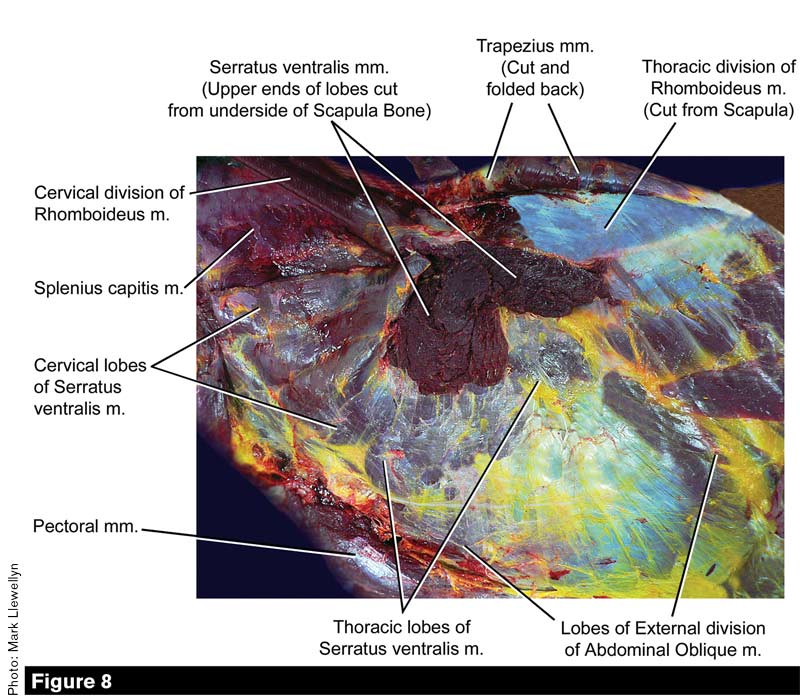
The same specimen under dissection, with bridging muscles marked.
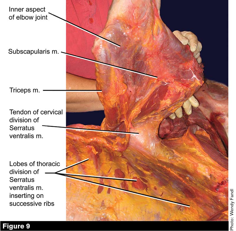
View of the inner aspect of the elbow joint and the undersurface of the shoulder blade. The animal’s head and neck extend to the right. The gloved hands support the shoulder (scapulo-humeral joint), while the right forelimb is lifted straight up in the air (the pectoral muscles were cut to make this possible). In lifting the leg so high, we bend the scapular cartilage, which is pressing against the withers and still attached to the DSL and trapezius and rhomboideus muscles. Note the extremely strong tendon of insertion of the cervical division of the serratus ventralis muscle that has been exposed by lifting the horse’s leg. The tendon of the thoracic serratus ventralis is partly visible just behind and deep to the previous.
Thoracic Sling
Despite its large size, multi-lobed structure and the strength of its attachment to the underside of the scapula and scapular cartilage, the serratus ventralis muscle is not the most important mover of the forelimb. Instead, the most important role of the serratus muscle is to form the major component of an elastic “thoracic sling” (Figures 5 and 6).
Understanding this begins with a significant fact: when they are not actively contracting, muscles work exactly like yellow ligaments. If you’ve been following this series from the beginning, you know exactly why: a muscle that doesn’t contract is – for all intents and purposes — a yellow ligament. To put this another way, muscles have a dual function: when actively contracting, they move the skeletal parts to which they are attached; but when not contracting, they act as passively stretchy “bungee cords.” If you only conceive of muscles as the source of bodily strength, you have missed 50% of the story, as they are also major mediators of bodily flexibility.
The lower, minor part of the thoracic sling is formed by the group of pectoral muscles (Figure 5). The pectorals sometimes also act as an elastic sling (Figure 6), but they act more often as actively contracting limb stabilizers and adductors.
Because the horse lacks collar bones, it can easily get into a habit of moving crookedly, with its sternum continuously off-center. This is, unfortunately, a tendency that the riding and training technique of many amateur owners and professional trainers exacerbates. Any tendency to one-sidedness in a given horse’s movement will quickly be reflected in asymmetrical muscle development and a tendency to one-sided soreness in the serrati and pectorals.
All the forelimb bridging muscles are prone to strain, so that very frequently the insertion of the latissimus dorsi, the lower third of the brachiocephalicus, the trapezii and rhomboids, and the pectorals become prime targets for chiropractic or massage therapy.
If the insertions of the serrati were more accessible in the living horse, I believe they would more often be overt therapeutic targets, because when the “lats” and “pecs” are sore, the serrati are sore, too. Likewise, changes in training or riding practices that benefit the more external bridging muscles also benefit the serrati.
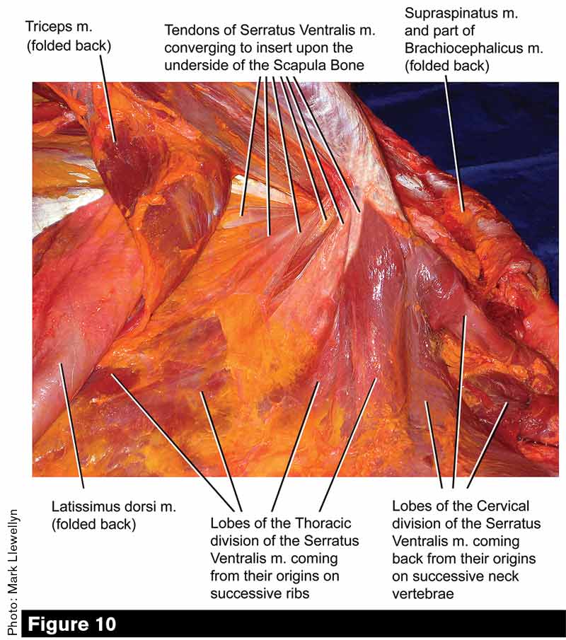
This view of a different specimen affords a good look at the tendon of insertion of the thoracic division of the serratus ventralis. Fibers from its several lobes converge to insert upon the periosteum on the underside of the scapula bone and the underside of the scapular cartilage.
This view looks down upon the specimen’s back from a position somewhat behind the shoulder. The gloved fingers are pulling the flexible scapular cartilage up to expose a thick padding of “bubble wrap.”

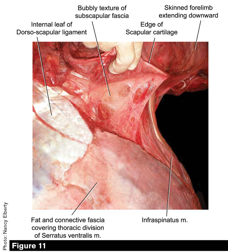
This view from the front represents a different approach to exposing the fascia underlying the shoulder blade; note its thickness and bubbly texture. The specimen is lying on its right side with its head extending toward the viewer. We have freed the anterior deep pectoral muscle from its attachment to fascia overlying the supraspinatus muscle on the cranial aspect of the shoulder blade. The brachiocephalicus muscle has also been cut from its attachment to the arm. Student assistants spread the horse’s forelimbs while I snapped the photograph.
Bubble Wrap Fascia
Another aspect of the junction between shoulder and rib cage that is important but seldom mentioned is the “heat sink” that pads the whole surface where the forelimb and thorax meet. In engineering, a “heat sink” is any structure or substance that absorbs heat since heat can be said to “sink” into it. For example, the radiator in your pickup truck — with its network of tubes filled with anti-freeze — is a heat sink that helps to keep the engine from working at too high a temperature.
The insulation in the walls of your house is also a heat sink, especially on hot summer afternoons. In the horse, a thick pad officially termed “areolar fascia” lies between the inner surface of the shoulder blade and the rib cage (Figures 1B, 11 and 12).
But let’s be clear about our terminology: the Latin “areolar” means “bubbly” and in a wet lab, when you get into this part of the carcass, the tissue looks and feels remarkably similar to the manufactured packing and insulating material called “bubble wrap.” As the forelimb is peeled away from the carcass, you can hear the characteristic crackling sound of popping bubbles.
It’s important to realize that in the living animal this bubble wrap tissue does not lie between the scapula bone and the rib bones. In the living animal, the underside of the scapula is covered by a slab-like muscle called the subscapularis, while the outer side of the rib cage is coated by the serratus ventralis and external oblique muscles.
What the bubble wrap is there for is to prevent the muscles on the opposing surfaces from rubbing on each other as the shoulder moves. This is because the generated heat would easily be enough to cook and kill those muscles. Equine bubble wrap is additionally invested with a certain amount of loose fatty tissue, which further reduces friction by making the padding somewhat oily and slick.
As to whether any considerable amount of heat is generated, there can be no question; everyone who has watched a five-gaited class for American Saddlebred horses or a park show class for Morgans, Arabians or Tennessee Walking Horses will have noticed the copious sweat that streams from the shoulder area of these competitors. Weighted shoes, high action and high energy act to increase the scope of shoulder movement and the amount of friction generated. Even ordinary pleasure horses generate considerable heat under the shoulder when trotting or cantering.


Post a comment
Report Abusive Comment