

Third in a Series
In this series, Dr. Deb Bennett examines the equine hind limb. In this installment, Bennett explores the stifle.
Farrier Takeaways
- The function of stifle-locking is to allow the horse to rest the muscles of its hindquarters and allow it to snooze in a standing position. The stifle may, however, unexpectedly lock during locomotion.
- Stifle unlocking is at least as important as stifle locking in permitting normal equine locomotion.
- Sticking stifles and full upward fixation of the PPC unit are more frequent in horses with “post-legged” conformation, but dis-coordination can cause it to happen in a horse of any build, even one with highly angulated hind limb conformation.
At first glance, the stifle joint appears to be a huge mistake — the worst-designed part of the musculoskeletal system.
Unlike the shoulder joint, hip socket, hock, elbow, intervertebral joints or the coffin joint, the articular surfaces of the femur and tibia, which face each other to form the stifle joint, do not closely correspond in shape. There is no neat ball-and-socket fit, no tight tongue and groove geometry and because of that the joint appears to have been “post-engineered” with a whole slew of auxiliary rims, pads, and straps that work to stabilize it and hold it together (Figures 1 and 2).
In terms of embryology and evolutionary history, the stifle joint’s design is indeed something of an “afterthought.” Lungfishes (Figure 3), have several modes of survival under conditions of drought, all of which have been observed in their living descendants.

A, anatomy of the equine stifle joint; left stifle in lateral view. The stifle capsule contains viscous synovial fluid. In this cadaver specimen, it has been injected with saline solution, over-inflating it to show its shape and volume. B, this synovium sample taken from a yearling draft horse cadaver measured 12 ounces (cup holds 16 ounces). Color code: tan = bone; grey = sesamoid; yellow = joint capsule filled with synovial fluid; light blue = tendon; dark blue = ligament; green = cartilage (“M” stands for “meniscus”); red = muscle.
When a pond begins to dry up and become stagnant, lungfishes can switch from gill-breathing to lung-breathing. If dry conditions persist, they can burrow into the mud and estivate for months, emerging when rains return. They also can emerge from the water. Using their forefins like oars to dig into the substrate, they can pull their bodies across mud until they find another, deeper pond. This is possible because the pectoral fins of lungfish are muscular and they are structured within by bones homologous to those in the mammalian forelimb.1
Importance of Limb Twist Location
A more detailed look highlights the fact that despite their ability to drag themselves across land, lungfishes are still fundamentally aquatic creatures for the bone in the lungfish pectoral fin that corresponds to the humerus has a straight shaft (Figure 3). In mammals, by contrast, the humeral shaft incorporates a strong twist that has the effect of changing the orientation of the palmar surface of the hand. In lungfish, the palmar surface of the fin normally orients inward toward the lateral surface of the body. When the fish crawls across a mudflat, it must do so by swinging the fin forward and then digging the trailing edge into the mud — because, with a straight humeral shaft, it cannot orient the palmar surface downward.2,3
Ancient amphibians were the first to develop a twisted humeral shaft that reorients the limb so that when it swings forward the palm can be set down flat against the substrate (Figure 4).3,4 Reptiles, birds and mammals, including horses, have inherited this design (Figure 5).5 Because the necessary twist is incorporated into the shaft of the humerus, the elbow joint and all the bones and joints more distal to it need none. The design of the elbow joint, for example, is very straightforward. It functions as a simple hinge that can make but one motion, flexion-extension — and, as a result, in horses and other mammals, the elbow joint is rarely injured during normal locomotion.

End-on view of the top of a left tibia showing the menisci and cruciate ligaments that are unique to the stifle joint. Anterior to top. The cruciate ligaments connect upward to deep fossae in the distal femur. They serve to stabilize the joint when the tibia rotates about its spine. Ligaments that anchor the menisci to the tibia (the menisco-tibial ligaments) are shown in purple.

There are 20 living species of lungfish, which have survived for millions of years. A, Australian lungfish Neoceratodus. B, the coelacanth Latimeria. The bone structure of the pectoral fin in each is shown at left. The element homologous to the mammalian humerus is highlighted in yellow.

The amphibian Ichthyostega lived at the end of the Devonian Period, about 360 million years ago. It was a fairly large animal, measuring 2 to 3 feet in length from nose to the tip of tail. Capable of crawling through muddy swamps, it possessed a twisted humeral shape (yellow) that permitted it to place its forefeet palm-down against the ground. By contrast, the shaft of its femur (green) is not twisted; neither had it developed a twist within the femoro-tibial joint (the equivalent of the equine stifle) so that the hind limbs were merely dragged along parallel to the body when it moved across land. Light blue color indicates restoration of missing elements. The carpal bones and tail vertebrae in this species remained cartilaginous throughout life, did not fossilize and are therefore not preserved in the fossil record. Abbreviations: H = humerus; R = radius; U = ulna; F = femur; T = tibia, Fb = fibula.

Humerus and femur bones of the horse in posterior (caudal) view, reduced to approximately the same length. The femur has a straight shaft, whereas the strong twist in the humeral shaft is easy to see.
The anatomical and functional changes that occurred through time in the vertebrate hind limb are, however, another story. In ancient amphibians, the better the twisted humerus worked, the less the need for the pelvic limb to assist in crawling — the hind feet could just be dragged along with the body (Figure 4).
Many other changes occurred over a long time before the fossil record presents vertebrates in which the palmar surface of the hindfoot can be set down flat against the ground; these animals include the reptile family from which mammals arose. The genes that govern the developmental sequence in these animals do not allow for a twist (analogous to that in the humerus) to develop in the femur (Figure 5). Instead, the necessary twist develops primarily within the stifle joint and it is this that necessitates all the ancillary reinforcement in the form of menisci and cruciate ligaments that we find in this joint.5
In a large animal such as a horse, fast running generates significant forces (torque, angular momentum, concussive, bending) that make it necessary for the hoofs to meet the ground “flat.” As part of the twist built into the stifle joint, horses have a prominent tibial spine that fits into a deep notch (the intercodyloid fossa) in the distal aspect of the femur (Figures 6 and 7). The spine allows rotation around the long axis of the tibia in addition to flexion and extension of the stifle joint.
In normal equine locomotion, the tibia (and all structures below it) rotates inward at the extreme of protraction, then rotates outward again as the limb draws back. As we learned in our last installment (see American Farriers Journal September/October 2021, Pages 52-60), the plane that bisects the horse’s hind limb orients outward when the horse stands still. As the limb is brought forward and the tibia rotates inward, the hind limb plane is brought parallel or nearly parallel to the midline plane of the body. This allows the hind hoof to strike on both heels at the same instant and allows the toe to orient forward rather than outward at breakover.

Bones that articulate to form the stifle joint. A, end-on view of distal end of the femur. B, end-on view of the proximal end of the tibia. C, tibia viewed from the front.

Digital X-ray of left stifle joint in lateral view; horse’s head to left. This view shows how the tibial spine (arrow) fits up into the space between the distal condyles of the femur.
Patellar “Ligaments”
Before the stifle locking mechanism can be properly understood, it is necessary to clear up another of the many conceptual confusions in equine anatomy study that have arisen out of traditional terminology.
In our first series, I pointed out that sesamoids, although they are ossified like bones, are not true bones because they do not have the same embryological development and they lack a periosteum. Sesamoids are intra-tendonal bursae that ossify (see American Farriers Journal December 2019, Pages 44-53). The patella is the largest sesamoid in the mammalian body. In the installment just cited, a detailed discussion concerning the nature of tendon tissue was also presented. Tendons differ from ligaments in terms of cellular composition and histological appearance, but also developmentally and functionally.
Every standard anatomy book refers to the three strong bands that connect the patella downward to the tibia as “patellar ligaments” (Figures 1, 11, 12). If a ligament is defined as a connective tissue band that connects bone to bone, then the use of the term “ligament” in this context is completely inaccurate since the patella is not a bone. Since the patella is, as stated above, an ossified intra-tendonal bursa, the patellar “ligaments” are actually the tendons of insertion of the quadriceps muscle and this is what embryological studies prove them to be.6,7 For this reason, throughout this article the patellar “ligaments” are referred to in quotation marks.
Snoozing vs. Deep Sleep
The stifle locking mechanism is a specialized hook-and-loop system possessed by equines (and, in a striking example of parallelism, also by certain specialized ruminants8). The anatomy that comprises the locking mechanism is not present in the earliest equids such as Eohippus or Miohippus. The enlarged medial epicondyle capped by a notch — the lockable stifle design — does not appear until the late Miocene in the genus Protohippus (in species extant about 12 million years ago). Protohippus, which was adapted to living in open savanna, is a direct ancestor of the modern horse genus Equus.8,9
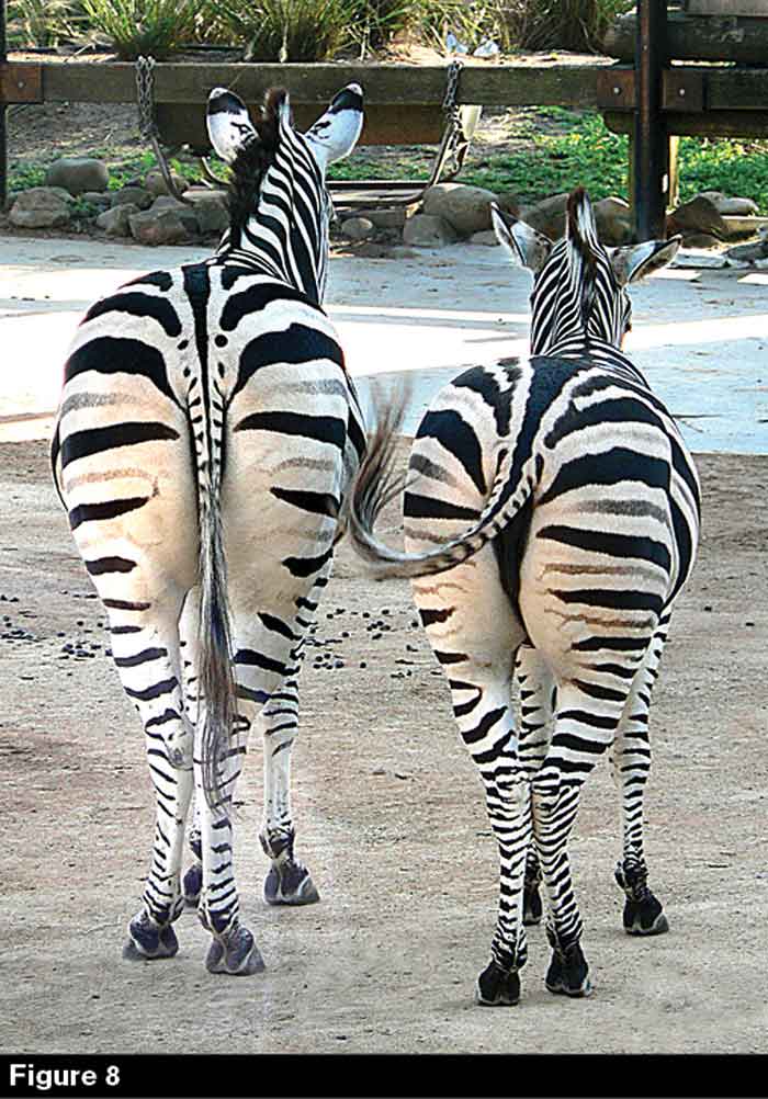
A pair of Burchell zebras having a companionable afternoon snooze at Taronga Zoo in Sydney, Australia. The zebra on the left has locked its right stifle joint and is resting its left hind leg; the one on the right has locked its left stifle joint and is resting its right hind leg. When an equine locks one stifle, the pelvis tilts over toward the opposite side; the striped pattern of zebra hide makes this very easy to see.
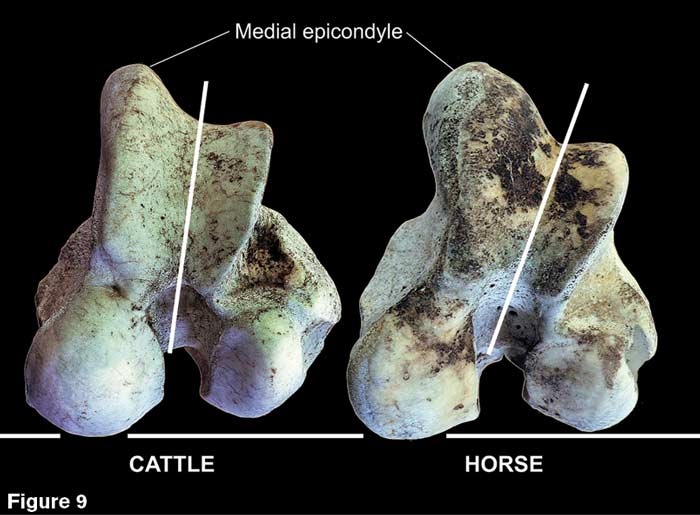
The distal end of the femur in cattle vs. horses. Differences in shape between the two species of large-hoofed mammal are subtle but significant. Both species have an enlarged medial epicondyle, but the epicondyles are more asymmetrical in horses than cattle (white lines). Cattle also lack the notch at the top of the medial epicondyle and are thus unable to lock the stifle.
The function of stifle-locking is to allow the horse to rest the muscles of its hindquarters and allow it to snooze in a standing position. To “rest a hind leg” the horse engages the locking mechanism on one side (Figure 8). The need for this is related to herd behavior as well as physiology. In deep sleep, mammals lose consciousness and along with that, control of most muscles; a horse that loses consciousness while standing will fall regardless of whether its stifles are locked. To sleep deeply, a horse must therefore lie down.10
Horses do not sleep straight through the night. The longest deep (rapid eye movement or “REM”) sleeps they take are in the range of about a half-hour. During the night, they periodically awaken, arise and graze. Nonetheless, horses require a total of 5 to 7 hours of sleep per day. Since they generally do not sleep through the night, they are fond of taking an afternoon nap. Typically, one herd member stands guard while the others lie down. The standing horse merely dozes and thus remains conscious. The stifle locking mechanism, which holds the locked hind limb in extension, allows him to doze, getting what is called slow-wave sleep.10 From one day to the next, “guard duty” is passed around the herd, so that every animal gets an adequate amount of rest.11

The anterior compartment of the stifle joint in this specimen has been opened. The capsule has been split open and most of the yellow synovial fluid allowed to drain out. The medial patellar ligament has been transected and the PPC unit swung open towards the lateral side like a gate, exposing the shiny bluish-gray hyaline cartilage that lines the opposing surfaces. The line of junction between the patella and patellar cartilage (dark red) is easy to see and palpate. The inferior rim of the PPC unit has been highlighted in purple; it is this rim that slides over the medial epicondyle “hook.”
Stifle Locking Mechanism
In many of their adaptations, cattle and horses parallel each other. Cattle have an enlarged medial epicondyle8 (Figure 9) but lack the notch that permits the equine stifle to be rigidly locked into the extended position (Figures 1, 11, 12). Cattle also do not characteristically “rest a hind leg” as horses do. Shape differences between the femurs of cattle and horses are subtle but significant. There is more asymmetry and outward orientation in the distal articular surface of horses than in cattle (white line in Figure 9).
The “loop” portion of the locking mechanism is created by the patella and patellar cartilage that is firmly anchored to it (forming the patella-patellar cartilage or PPC unit) (Figure 10), plus the patellar “ligaments” that tie the PPC unit down to the tibia. The inferior rim of the PPC unit plus the medial patellar “ligament” form a smooth, broad “U” shape (similar to that formed when you close your fingers but spread the thumb wide to form the letter “C”). It is this edge that lodges in the notch atop the medial epicondyle to lock the stifle joint.
One can easily observe the horse deliberately lock one stifle to rest a hind leg; contraction of the quadriceps muscle that lifts the PPC unit is easy to see. The quadriceps then relax, dropping it into the notch above the medial epicondyle. The animal stands higher on the locked hind leg with the hips rolled over toward the opposite hind limb, which often will be flexed and rested upon the toe (Figure 8). Because the patellar “ligaments” are strong but not very stretchy, when the PPC unit lodges in the medial epicondyle notch, the stifle joint cannot be flexed (Figures 1, 11, 12).

The stifle locking mechanism; a right stifle in lateral view. Above, unlocked position; below, locked position. The color code is the same as in Figure 1a.

The stifle joint locking-unlocking mechanism as seen from the front. From extension (yellow arrow), the patella can be pulled upward and then released behind the medial epicondyle; this is the fully locked position (red arrow). In movement, the TFL muscle must fire at exactly the right instant; the normal sequence is from extension (yellow) to unlocking to flexion (green arrow). When the flexor muscles contract (curved purple arrow), they pull the distal end of the tibia caudally and the patellar “ligaments” pull the patella downward in the patellar groove.
Significance of Unlocking
During normal flexion and extension of the stifle, the patella slides up and down in the patellar groove. The quadriceps pull the PPC unit upward when the joint is opened and the patellar “ligaments” pull it downward when the flexor muscles pull the tibia back to close the joint (Figure 12). The tensor fasciae latae muscle (TFL) is the most external of the muscles that extend the stifle joint. Because of its origin on the point of the hip, it is crucial not to stifle locking but to the even more necessary stifle unlocking (Figure 13).

The LaBrea Tar Pit horse, Equus occidentalis, skeletal mount in rear view (on display at the George C. Page Museum in Los Angeles, Calif.). The TFL muscle (in red) is the only hip flexor/stifle extensor whose origin is so far away from the midline plane of the body. Its lateral position gives it leverage so that when it contracts it pulls the patella up and laterally and then holds it up long enough for flexion of the joint to begin (see Figure 12).
The stifle joint is opened wide twice in every trot stride (camera frames marked by red stars in Figure 14) and may be opened so wide that the PPC unit is pulled high enough to clear the medial epicondyle and thus potentially catch on the femoral hook. This is the case even in horses with excellent conformation and even in riding styles that call for sharp flexion of the hind joints. This constitutes a serious downside since having the locking mechanism unexpectedly engage during locomotion jerks the horse to an abrupt and painful halt. If the horse is moving at any speed, it can tear or catastrophically rupture the patellar “ligaments.”12
It is also possible for the locking mechanism to partially engage without fully locking (“delayed patellar release”12). When this happens, the PPC unit drags over the top of the medial epicondyle (“sticking” or “rubbing” stifles). This irritates or can even abrade the inner lining of the PPC unit, provoking fluid buildup and swelling (a common cause of gonitis13) (Figure 15b).
Sticking stifles and full upward fixation of the PPC unit are more frequent in horses built with wide-open hindlimb joints (“post-legged” conformation) (Figure 15a), but dis-coordination can cause it to happen in a horse of any build, even one with highly angulated hind limb conformation.
The TFL, the muscle responsible for unlocking the stifle and for preventing it from becoming locked during locomotion, is structured with a relatively thin belly overlying a long, flat tendon. Its contraction is only just sufficient to raise the PPC unit above the medial epicondyle. It is crucial therefore that the TFL contract within a small window of time — a matter of milliseconds — at the two points in the trot stride (full protraction and full retraction) during which the PPC unit has been lifted by the quadriceps high enough to catch.
The job of the TFL is to raise the PPC unit above the medial epicondyle hook at just the right moment and then hold it there for long enough to permit flexion to begin (Figure 12). The TFL then relaxes and the PPC unit is pulled downward in the patellar groove as flexion of the joint continues.
A little calculation highlights how important it is that this sequence occurs on time, every time with each stride and how small the window is for effective TFL contraction. A moderate trot proceeds at about 80 beats per minute, that is, either “clip” or “clop” will be heard about once every three-quarters of a second. If we consider only one hind limb, that limb will cycle from full protraction/extension to full retraction/extension 40 times per minute, and the stifle joint will thus be fully opened in that limb 80 times per minute. This means that the TFL in each hind limb needs to contract once every three-quarters of a second for so long as the horse continues to trot.
Rather than asking why the stifle joint occasionally “catches” in some horses, on this basis, it would seem more reasonable to ask why it does not catch more often. The answer lies in the horse’s amazing neuromuscular integration, which allows for the precise coordination of the firing of the quadriceps, TFL and flexor musculature as pictured in Figure 12.

Sequence frames of a Morgan park horse trotting. This style of movement requires more flexion of the hind joints than is typically seen in dressage, hunter or Western pleasure. Despite this, the hind limb joints must still be opened twice in every trot stride (red star in frames 5 and 8). At these moments, the patella may be pulled high enough to rise above the medial epicondyle and thus could potentially lock.
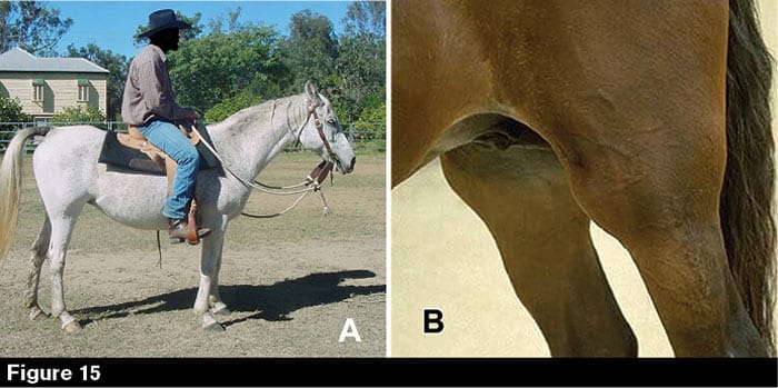
A, an Arabian mare with “post-legged” conformation, in which the stifle and hock joints are open to nearly their full extent when the animal stands. Horses with this conformation can be prone to delayed patellar release and/or upward fixation of the patella. B, chronic delayed patellar release, often called “catching,” “rubbing,” or “sticking” stifles, irritates the cartilage lining of the PPC unit, resulting in fluid accumulation and swelling of the joint (gonitis).
References
- Jarvik E. 1996. The Devonian tetrapod Ichthyostega. Fossils and Srata 40:1-206.
- Ahlberg PE, Clack JA, and Blom H. 2005. The axial skeleton of the Devonian tetrapod Ichthyostega. Nature 437:137-140.
- Sillezar J. 2020. From fins to limbs and water to land: Harvard scientists reconstruct the evolution of terrestrial movement in early tetrapods. The Harvard Gazette (available at news.harvard.edu/gazette/story/2020/11/reconstructing-vertebrates-rise-from-the-water-to-land).
- Hildebrand M. 1974. Analysis of Vertebrate Structure. Wiley, New York, 710 pp.
- Romer AS. 1977. The Vertebrate Body. W.B. Saunders, Philadelphia. 679 pp.
- Gardner E and O’Rahilly R. 1968. The early development of the knee joint in staged human embryos. Journal of Anatomy 102:289-299.
- Tria AJ, and Alicea JA. 1995. Embryology and anatomy of the patella, in: Scruder, G.R. (ed.) The Patella. Springer, New York, pp. 226-298.
- Hermanson JW and MacFadden BJ. 1996. Evolutionary and functional morphology of the knee in fossil and extant horses (Equidae). Journal of Vertebrate Paleontology 16(2): 349-357.
- Bennett DK. 1992 (revised and expanded 2005). The evolution of the horse: history and techniques of study, in: Evans, J.W. (ed.), The Elsevier World Animal Science Encyclopedia, vol. C7 Horse Breeding and Management, pp. 1-37 (available online at equinestudies.org/knowledge_base_intro/evolution_pdfs_intropage.html)
- Burla JB, Rufener C, and Bachmann I. 2017. Space allowances of the littered area affects lying behavior in group-housed horses. Frontiers in Veterinary Science 4:23 (available at kentuckyequineresearch.com/equinews/sleep-requirements-horses).
- Berger J. 1986. Wild Horses of the Great Basin: Social Competition and Population Size. University of Chicago Press, 246 pp.
- Boswell JE. 2015. Intermittent upward fixation of the patella and delayed patellar release in horses. Merck Veterinary Manual (available at merckveterinarymanual.com/musculoskeletal-system/lameness-in-horses/inter).
- Boswell JE. 2019. Disorders of the stifle in horses. Merck Veterinary Manual (available at merckveterinarymanual.com/musculoskeletal-system/lameness-in-horses/inter).
- Sisson S, and Grossman JD. 1953. The Anatomy of the Domestic Animals. W.B. Saunders, Philadelphia, 972 pp.


