It’s the early hours of Derby Day Minus One as I arrive at Rood & Riddle Equine Hospital in Lexington, Ky., a time when the eyes of the world turn to the Bluegrass Country more than any other time of the year.

But while there is plenty of Derby talk and chatter, the horses that will run in the Derby and its related races number only a few score, and there are thousands and thousands of Thoroughbreds in the Bluegrass. The attention of the sporting world may be focused on Churchill Downs, but there’s plenty of work that still needs to be done by those in the equine industry — Derby or no Derby.

Raul Bras, a veterinarian and certified journeyman farrier who works in the Podiatry Department at Rood & Riddle, is preparing for a full day of providing hoof care both at the hospital and at area barns. As we’ll see, Shoeing For A Living presents a broad range of challenges for those charged with keeping Thoroughbreds sound.

7 a.m. Bras and his assistant, Josh Wilbers, have already made sure their rig is fully stocked for the morning’s barn calls. But before they head out on the road, they have some work to do with a high-end Thoroughbred colt. The horse was brought to the hospital two days earlier, after it developed pneumonia. The stress of the illness, combined with the colt’s negative reaction to being trailered to the facility, resulted in the colt displaying some warning signs of laminitis.

The attending veterinarian called in the Podiatry Department. Finding the front feet hot, and what appeared to be elevated pulses, Bras had the horse placed in ice-filled boots.

7:08 a.m. The colt has spent the last 24 hours with his feet in those boots in an effort to head off the laminitis. The colt is brought out from the stall into the clinic aisle. The thick, blue plastic boots that hold the ice slurry are removed, as are smaller hoof boots worn inside the ice boots. Bras checks the pulses on all four feet, and also palpitates the coronary bands to see if they are sinking. He finds the pulses are much lower than they were before the ice was applied — an encouraging sign.

Bras does some light rasping around the heels of the hoof walls, explaining that he’s “just roughing up the heels a bit to give him a flat surface to stand on.” He has to be very careful, as the colt has what he described as “typical racehorse feet — no walls and very thin soles.”

Bras removes a treatment plate to check on how a surgical site is healing. Sterile maggots are being used to keep the site clean of bacteria. Once he’s satisfied with the progress, he replaces the treatment and bandages the foot with a combination of Elastikon and duct tape to protect the foot from mud, manure and other debris.

Bras traces a hoof on a piece of paper at a Lexington-area Thoroughbred farm. The tracing will serve as a template for building a shoe back at the Rood & Riddle farrier shop. Bras and Wilbers will return the following day to apply it.

Wilbers first replaces the hoof boots, followed by the ice boots, which are then refilled with ice. Bras points out that one of the tricks to using the ice is to make sure it doesn’t all wind up at the bottom of the boot.

“He’ll try to pick his feet up and stand on all the ice. I want it to go all the way up to his knees, so you kind of have to manage it,” he explains as he and Wilbers work together to get the ice distributed within the boots the way he wants it.

Bras is pleased with how the colt is progressing. When he was first placed in the boots, he didn’t take to them very well.

“He was having some trouble figuring them out and getting used to them,” Bras says. “We were worried that he was getting a little stressed out with the boots. We think he developed his original problems due to stress, and we definitely didn’t want to add to his stress.”

But this morning, the colt seems calmer. Bras notes that he’s resting his feet, not constantly shifting his weight from one to the other or leaning back on his rear haunches, as horses will when suffering extreme discomfort for laminitis.

“He’s much better,” Bras says. “I’m trying to read the whole horse here, and I can see that he’s got his head up, his eyes are bright, he’s checking out things around him. Yesterday, we could hardly get him to walk out of the stall.”

Bras observes a foal with its dam. The foal was born prematurely and is being treated for contraction in its hind legs. Note that the foal is still standing on its toes.

Bras talks with Dr. Stephen Reed, the attending veterinarian, and recommends that the colt be kept in the ice boots for three days.

“This type of case can be one when you are damned if you do and damned if you don’t,” he says as the horse is led back to his stall. “If you take him off of this kind of treatment and he ends up foundering, you’re in pretty bad shape.”

Bras says that if a high-level racehorse founders, owners are often done with them. If this colt recovers, without any rotation or sinking, his prognosis for a return to racing is favorable.

“Things get really tricky when we take the boots off and the feet begin to reperfuse with blood. Then we really have to keep an eye on him,” he says. “We have to follow him, make sure he’s got some support and that he doesn’t start having new troubles as the triggering factors from the underlying disease can reach the circulation in his feet.”

So far, radiographs have not indicated any rotation. Bras says this is a case where it appears they have been able to start treating the potential laminitis at a very early stage.

“We can’t prevent what’s already happened, but we can try to prevent further damage,” he says. “Laminitis is often secondary to the primary disease, as is the case here. If we can get his primary disease taken care of, hopefully we won’t have any more insult to his feet.”

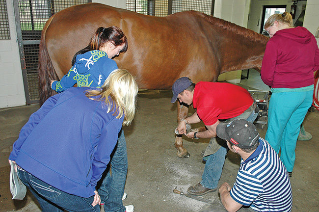
After sedating the foal, Bras and Wilbers are joined by a trainer in applying splints to the hinds. The splints are made from 3M casting material.

Bras says later today, the feet will be radiographed and the results compared to those taken the previous day. The hoof boots the colt wears inside the larger ice boots are set up to transfer weight off the walls and to share weight bearing with the sole to support the bony column.

“These are the most frustrating cases to work with, but they are the most rewarding as well,” Bras says as he pilots the rig down the driveway toward the highway. “If they come around, you really feel good.”

8:46 a.m. The first barn call of the day is a follow-up call on a yearling filly that had undergone surgery earlier to correct a conformational problem.

“We spend a lot of time working on conformation,” he says. “Obviously, we look at a lot of Thoroughbreds before they go to the sales. They have to look as close to perfect as possible.”

The splints are then tightly bandaged with Vetrap. Bras next uses his T-square in an unusual way. He inserts it through the bandage near the toe, then lays it across the toe and uses it to apply leverage, stretching the contracted tendons.

This particular filly’s surgery was to correct a fetlock varus condition, with the knee offset from the cannon bone. Bras explains that she has been shod with shoes forged to help build up some outside flare on her front feet for a more normal appearance.

The barn manager mentions that he’s anxious to see how the horse moves when the shoes are removed, noting that the screw is still in place in the knee, but that the alignment of the knee looks much better.

8:51 a.m. Wilbers removes the front shoes, despite the fact that the filly is not very cooperative.

“I’m kind of letting Josh get beat up,” Bras says. “But I don’t want to sedate her, because it’s important to see how she walks without the shoes. Sedating her would affect that.”

A groom walks the horse up and down the barn aisle after the shoes have been removed. Bras and the barn manager agree that the filly is showing considerable improvement and appears to be on the right track for the September sale. He notes that they will want to encourage a little more flare on the left foot as opposed to the right.

He does some touching up of the front feet with a rasp, then sets each foot down on a piece of paper he’s placed on the barn floor. He traces the outline of the hooves on the paper to serve as a template for building the filly’s next pair of shoes in the Rood & Riddle shop.

9:13 a.m. Bras decides that they’ll wait to trim the hinds until a scheduled visit the next week.

“I don’t want Josh to get beat up too badly since she’s being such a riot,” he says. “Next time, we can sedate her and get the hinds trimmed up.”

9:20 a.m. During the short drive to the next stop, Bras handles a couple of cell-phone calls, updating Scott Morrison, the head of the Rood & Riddle Podiatry Department on the progress of various cases, and also talking to a barn manager. Throughout the day, he’ll spend a lot of time on the phone between barn calls.

9:34 a.m. A couple of horses are on the docket at the next barn; a mare being treated for complications from a chronic abscess, and a foal with ballerina syndrome caused by contracted tendons.

9:38 a.m. Bras checks the mare first. He explains that the problems started with a puncture wound near the frog.

“Those are the ones that can become a real problem, because they can involve the navicular bursa,” he says.

Although the wound was treated, it never really healed. Bras was called in after the mare developed the abscess.

Bras shoots radiographs of a foot with a suspected seroma and is quickly able to show the results to the barn manager.

“We took a radiograph and found that it had a track that went to the wings of the palmar processes of the foot,” he says. “There was some necrotic tissue, the bone was involved and there was some infection. That’s probably why she kept having abscesses and why the wound wouldn’t close.”

Surgery involved scooping out necrotic tissue, as well as cleaning up the bone with a curette.

Bras examines a hoof crack on a Quarter Horse gelding that is used for jumping. The crack, which Bras believes may have been caused by a medial lateral imbalance, runs all the way through to the sensitive inner tissues. The crack isn’t healing because granulation tissue that is forming gets pinched in the crack as the hoof expands and contracts.

“We also do local and systematic antibiotics,” he says. “We do a regional perfusion (using a tourniquet to concentrate the antibiotics in the area of infection) over three days and soak the foot in CleanTrax once the surgery site starts healing.”

The foot is also undergoing maggot therapy, in which sterile maggots are placed in the wound. Most often, maggot therapy is used because the maggots remove necrotic tissue. But in this case, Bras says the maggot secretions are antibacterial and are intended to keep bacteria out of the wound, as the necrotic tissue was removed during surgery. The foot is protected and the maggots kept in place with a treatment plate covered by a bandage.

9:47 a.m. During his initial examination, Bras finds that the horse has torn through the bandage, which is also covered in mud. He chides a groom for letting this happen, although upon opening the hospital plate, he finds that the maggots are still active.

Susan, the trainer of the horse, has come over during his inspection and he tells her the surgery site is coming along nicely. He also informs her about the damaged bandage and tells her the foot should be rewrapped if it happens again.

He replaces the cover on the hospital plate and applies a new Elastikon and duct tape bandage. Once the mare is back in her stall, Susan accompanies Bras and Wilbers to a nearby round pen, where the foal with the ballerina stance is being kept with her dam.

10:07 a.m. Bras is actually pleased to see that this foal is outside.

“I’m a little surprised that this foal is even standing,” he admits. “The first time we worked on her, she couldn’t even stand on her own.”

The foal was born prematurely with contracture in all four limbs.

“She was a complete train wreck when she was born,” says Susan.

As they observe the foal, Bras notes that while her front limbs have a near normal stance, she continues to stand on her hind toes.

Bras shapes a keg shoe to fit the foot. He fits it with a heart bar insert, which is then welded in by one of the farriers who works in the Podiatry Department shop. Bras is using the heart bar shoe to unload the hoof crack and transfer weight to the frog and other sound structures.

“I wouldn’t mess with her fronts at all right now,” he tells Susan. “But we need to do something with her hinds.”

Once the heart bar shoe was fastened with a combination of nails and glue, Bras had the horse wait in a stall for a few minutes. In a very short time, the jammed up wall above the crack had dropped so much that he needed to cut through the glue and use a Dremel tool to refloat the crack.

Bras cauterizes the soft tissue within the crack to keep it from getting pinched while the crack grows out.

Bras says he thinks that the hind-end contracture was the original problem and that the front-end contracture may have been, in part, a result of the foal compensating for that problem.

Susan and Wilbers control the foal. The mare looks on, but doesn’t interfere. Bras sedates the animal and she’s quickly laid down on her side in the pen. A towel is put over her eyes to help keep her calm.

This case presents a real challenge, Bras admits.

“There’s a debate about what causes these cases,” he says. “Laxity seems to happen more often with premature foals due to a lack of growth in the uterus, but contracture is harder to explain. Is it how they are placed in the uterus or some kind of issues that the mare had during gestation? Genetics? Nutrition?”

He says there is also a question about whether contracture and laxity involves the tendons or muscles. This foal also has some additional factors that complicate the case.

“If a foal has contracture, you want it to get some stall rest so that the affected areas will weaken and relax,” he says. “But in this case, you also have a foal that’s back at the knees. You have to have her get out so she’ll get stronger, because if she doesn’t, she’ll crush her knees. So we have to let her out (of the stall) so that the front gets better, but we have to be concerned that we’re overdoing it, causing the back end to contract again.”

10:13 a.m. Once the foals is quiet on the ground, Bras and Wilbers apply 3M VetCast Plus, a casting tape that is dipped in water before it is wrapped around the limb, where it will eventually harden. Before that happens, Bras takes his T-square and slips it through the end of the bandage and cast near the toe. He uses the T-square to apply leverage to the toe and stretch the tendon. The goal is for the combination of the hardened cast and bandage to hold the tendon in its stretched form.

Bras injects stem cells into the lower limb of a Saddlebred mare recovering from laminitis. Bras says that stem cells used in this way lead to accelerated growth. He hopes the therapy will help damaged laminae regenerate and strengthen their bond to the hoof wall.

“We’ll come back tomorrow and stretch it some more,” he says.

Vetrap is wrapped over the cast. Bras instructs Wilbers to wrap the leg more tightly as the bandage reaches the bottom of the foot. The towel is removed from the foal’s eyes. She blinks and looks about, but for now, seems content to recline in the warm spring sun.

10:36 a.m. Once it’s clear that the foal is coming out of the sedation, Bras and Wilbers pack up their rig and head for the next stop on the schedule. There are two mares that need to be checked at this barn. The first displayed signs of laminitis after foaling. She’s been shod with glue-on shoes. Bras checks her pulses and soles and concludes that she seems more comfortable than she was.

He says he’d actually like to replace the shoes, but doesn’t think the mare would stand for it.

“She’s a very, very heavy mare with small feet,” Bras explains. “She doesn’t really have the base of support she needs for her weight.”

The second mare is chronically laminitic. Bras wants to remove her current shoes and replace them with Sigafoos Series I SM shoes with a rocker toe. He’ll also use an Equi-Pak pour-in pad with the shoes.

“In these cases, it’s really important to do a good prep,” he says after the shoes have been removed. “I want to dry the sole out and get rid of any old, exfoliated sole. This sole has a lot of cracks, holes and crevasses. Fungus and white line disease will take advantage of that.

“We need to clean it up really good and try to kill any fungus that’s present. I’m going to cover it up with glue again, and if I don’t do my prep work right, I can actually make things worse for the horse instead of helping her.”

Bras passes a lighted propane torch quickly over the sole of each foot as part of the prep work. He’s careful in using the flame, which he hopes will kill the fungus and help remove any exfoliated sole.

“If a horse has a really tough sole, running a torch over it can make it a little easier to work with and loosen it up,” he says. “But that’s not what we’re trying to do here. She has thin walls and flat feet. I didn’t pare the sole during cleanup because she doesn’t have much. If you have a very thin sole like this and aren’t very careful with the torch, the horse will feel it.”

11:16 a.m. Bras also treats the sole and frog area with a little copper sulfate before applying the shoes. Wilbers uses Vetrap and tape to protect the coronet bands from the glue that impregnates the cuffs of the Sigafoos shoes. Once the shoes are in place and the glue has set. They glue foam boards across the soles of the shoes and inject the Equi-Pak.

11:32 a.m. Wilbers goes to work on the hinds once Bras is satisfied that the front shoes and pour-in pads have had time to set-up and will remain securely in place if the mare moves too much while her hinds are being worked on.

Wilbers has been riding with Bras for the past 10 months, after spending the previous 4 years working as a late shift vet tech at Rood & Riddle. In effect, he’s getting a shoeing apprenticeship under the tutelage of Bras and the other farriers and vets who work at the hospital. A couple of years earlier, he says he’d had an opportunity to take a similar position under another veterinarian/farrier at the hospital, but hadn’t been able to because he was needed to work on his family’s farm during the day.

“My family is in the process of selling the farm, so I’m not needed there as much any more,” he says. “I always regretted not being able to take advantage of that earlier opportunity, so when Raul gave me a second chance, I jumped at it.”

11:48 a.m. The next stop was added to the original schedule after Mark, a trainer and barn manager, called. Bras is asked to check on a filly that is 3 weeks into a 2-month layoff from training. The filly had been going barefoot, but had suddenly shown signs of lameness. Mark explains that he found a bruise in the sole, and then noticed blood. At first he suspected an abscess. He opened the sole in the affected area and got some drainage. He then decided to ask Bras to look at it.

12:06 p.m. Bras conducts a preliminary examination of the foot and immediately decides he wants to see radiographs before he goes any further. He’s worried about infection, noting, “If you get infection, you’ll get more problems.”

As he sets up for the X-rays using a portable digital machine, he explains that he’s worried about a possible infection of the coffin bone. The horse has a very thin, flat sole. Bras thinks a seroma may have developed beneath the sole.

While most of his day was spent working on Thoroughbreds, Bras also found the time to trim up the feet of a miniature horse, and drain and treat an abscess.

Bras and the horse’s owner look on during an ultrasound on a Saddlebred mare. The ultrasound uncovered a small tear in a suspensory ligament.

After examining the radiographs, Bras concludes his tentative diagnosis was right. He says the horse probably stepped on a stone, and the resulting bruise led to the seroma that has now become infected.

He also points out that the horse is a little long in the toe. That has the effect of stretching the sole, making it even thinner.

“I’d like to take off that long toe, open up the seroma a little to drain it, treat it and cover it with a treatment plate,” he says after showing the radiographs to Mark. “But if we back that whole toe up, we might lose the white line for nailing.”

Bras says they will put shoes back on the horse, make a tracing of the foot and come back the next day with a treatment plate to put over the shoe.

12:32 p.m. He has Wilbers trim the toe back just a little. He tells the trainer to soak the foot in Epsom salts, then pack the area with square cotton and Betadyne, adding that he’ll leave the necessary materials.

12:41 p.m. There is another filly Bras needs to look at. This one had splint bone surgery a short time before and has been turned out for the last couple of days. She started acting as if one foot was a little sore. When Mark examined the foot, he probed the heel and got a little pus. Again, with a thin-soled horse, he didn’t want to do too much digging.

As with the previous horse, Bras says the apparent infection is in “a rough spot,” where it could easily involve the hoof’s sensitive inner structures. He uses hoof testers and with just a little pressure, gets a reaction from the horse, as well as some additional drainage.

He cleans off the hoof a bit more and locates a small puncture. Mark says he’d like to bandage it and treat it with CleanTrax. He thinks the abscess would eventually erupt at the coronet band.

Bras agrees to that, but says the filly should be watched closely. He also says the foot should be soaked in Epsom salts. He agrees to leave some Animalintex pads for the treatment.

1:02 p.m. Over lunch at a nearby barbecue restaurant, Bras talks about both of the cases.

Bras checks the foot casts of a mare that’s being treated for laminitis. Throughout the day, he’s frequently on his cell phone, keeping in touch with the hospital, as well as other veterinarians and clients.

“With the first horse, there was radiographic evidence of roughening of the solar margin of the coffin bone, as well as pedal osteitis,” he says. “She’s been in the barn a couple of weeks and came in a typical race horse — low heels, long toes, thin soles.”

He says these problems are complicated by the fact that racehorses are shod more frequently than most horses. The soles often wind up being pared down a little each time. Add that to a conformation that may make it difficult for the horse to grow sole to begin with, and you’re headed for trouble.

“We have to deal with the issues we have right now, but also look down the road,” he explains. “We can’t put too much pressure on the sole, but we have to come up with something to give some stimulation of that sole. If it has stimulation, it will grow stronger.”

When they return the next day, they will shoe the filly with a Sigafoos Series I, combined with a treatment plate.

“We’ll just load the back part of the foot and pack it with cotton. We want to keep it dry. If Mark feels like the tissue starts to dry, I’ll tell him to use Keratex so it will dry and cornify.”

He says he was a little surprised that the X-rays showed that the coffin bone wasn’t flat or negative in alignment, as he often sees in long toe and low heel syndrome.

“With that, you often slow down the growth of sole depth due to compression on the circulation on the solar plexus,” he says. “But she wasn’t bad. She was still tipped pretty good.”

Bras says that his goal with the shoe will be to get the sole up off the ground a little and incorporate the foot’s stronger structures for support, primarily the frog in this case.

Bras will initially fill the sole with cotton soaked in a mild astringent to stimulate sole growth.

“I won’t use Keratex at first because I don’t want to burn the tissue,” he explains. “But once we get it healed up and built up a little, we’ll pack it with Keratex. In 5 weeks when we shoe her again, I won’t even touch the sole. Whatever I can grow, I’ll keep. She’ll still be flatfooted, but at least she’ll have some protection.”

Bras says sometimes trainers want farriers to fix a flat foot by making the sole concave. This results in an even thinner sole, sometimes dangerously so.

“We’ve had some cases where the sole is so thin, you get a little hole and there will be sensitive tissue coming out through the hole like a cauliflower,” he says. “It won’t heal because that sensitive tissue is getting pinched as the hoof expands and contracts. That can lead to an infection of the sensitive tissue. I have to cauterize the area in cases like that.”

After completing veterinary school, Bras, a native of Puerto Rico, completed a surgery internship at Rood & Riddle. He then apprenticed under Scott Morrison, the vet/farrier who leads the Podiatry Department, followed by yet another year shoeing sport horses for Rood & Riddle. He then went to Cornell, where he completed the farrier program being taught by Mike Wildenstein, the since-retired member of the International Horseshoeing Hall Of Fame. Finally, he returned to Rood & Riddle.

1:55 p.m. After lunch, Bras and Wilbers return to the hospital. They’ve been called in to look at a 6-year-old Quarter Horse that was trailered down from near Columbus, Ohio.

The horse is a jumper and his owner is hoping to ride him in a competition on Sunday — just two days away. But the horse also has a bleeding quarter crack. After an initial look, Bras doesn’t think making the show is very likely.

“I will let him do it if he can,” he explains to the horse’s young owner. “But not if he’s going to be lame for a long time after that as a result.”

The owner assures Bras she doesn’t want to risk injuring the horse and he continues with his exam. He asks questions as he watches the horse walk, picks up the feet and shoots radiographs.

“My concern is the bleeding,” he says. “I could put a patch on the crack, or stitch him up, but the bleeding indicates we’re already into sensitive tissue. It looks like there might be a little infection. If I cover everything up, I can create a bigger problem.”

2:17 p.m. The crack begins to bleed anew, at just the lightest touch of a rasp on the foot. After looking at radiographs and taking a close look, Bras concludes that the crack has indeed penetrated into soft tissue. Granulation tissue is forming in the area, but the wound is not really getting a chance to heal.

“When he loads, this closes,” he says, demonstrating the motion of the crack with his hands. “When he unloads, it opens. So when he jumps, then lands, he pinches it, and pinches it and pinches it,” he says.

He’s also decided that the horse has some balance issues.

“What’s really important here is not necessarily patching it or fixing it, it’s sorting out why your horse is developing a crack,” he says. “A crack is usually secondary to something else; a conformational deformity or the way the horse has been trimmed or shod. These are things that can create an imbalance.”

Bras explains that he wants to make sure the horse has a solid base of support. He says that may involve taking weight off the crack and transferring it to the frog and other stronger areas of the hoof capsule.

“We’ll see what we can do, as well as what he’ll allow us to do, and try to come up with a shoe that will help him,” he says.

Bras says one problem is that the lateral heel on the right front is diving in, leaving no room to nail without going inside the white wall.

“If I put nails in him, I’m jeopardizing him and taking a risk of pricking him or worse,” he says, adding that’s probably why the previous farrier had used glue in the area.

2:32 p.m. Bras tells the owners that he has a plan for the horse, but he’s also frank about the cost, telling them this isn’t a simple shoeing job and will be expensive.

“I will try to do as much as possible at the best price I can,” he tells them. “If I can give you a break on a couple of things, I will.”

The veterinarian also says the horse won’t be able to compete at the Sunday show — or anytime soon, for that matter. He believes he can remove the stress that is causing the crack and that it will grow out, but says the process will probably take at least two or three shoeings.

“I’m sorry we’re giving you such bad news,” he says. “We can help him, but it’s going to take some time. You’re not going to be able to go riding on him for a while. He can’t be jumping. He can be hand-walked, but it will have to be a very slow walk. If he overdoes it, it will defeat the whole purpose of what we’re trying to do.”

Bras decides on a heart bar shoe to unload the crack. He thinks part of the horse’s problem is a medial lateral imbalance that has pushed the wall up, causing stress that eventually led to the crack. He says he’ll also cauterize the crack to keep the tissue from getting pinched.

3:32 p.m. Bras carefully trims all four of the horse’s feet, then shapes and fits a steel shoe for the foot with the crack. He doesn’t feel aluminum would be a good choice for the big Quarter Horse. He also points out that one of the somewhat tricky aspects of this shoeing is that one of the front feet requires an advanced therapeutic shoe and the other doesn’t. But he has to “balance” the fronts to a certain extent. He doesn’t want one shoe to be significantly heavier than the other, or raise one side of the horse higher than the other.

3:35 p.m. Once he’s satisfied with the shoe’s fit, Bras turns over welding of its heart bar insert to one of the farriers who works in the shop. Wilbers uses a buffer to clean up the hinds. Meanwhile, Bras turns his attention to a Saddlebred mare.

Bras has been treating this mare for laminitis. She had some rotation that was made worse by a bad case of white line disease. The horse has made a lot of progress and has actually returned to work. But while observing the horse during a training session earlier in the week, Bras noticed she was still favoring the right front.

“She’s doing pretty well, but she’s still not quite 100%,” he says. “This is the kind of horse that wins the whole thing when she goes to a show. We really want to get her back to that level.”

The mare has been brought in for a couple of treatments. Bras plans to inject the recovering foot with stem cells and will also have an ultrasound done to see if there might be an injury in a tendon or somewhere else on the limb that might be contributing to the lameness.

Veterinarians at Rood & Riddle have used stem cell therapy in a variety of ways, according to Bras. The equine hospital harvests its own stem cells for use. In this case, Bras injects the stem cells just above the hoof, using a tourniquet above the injection site to concentrate the stem cells in the area.

Bras hopes the therapy will help regenerate laminae that have been damaged, as well as promote a stronger bond between laminae and the hoof wall.

“We really don’t understand just how stem cell therapy works, but we’ve found that with them, we get a lot of hoof growth,” he says. “When you get a lot of growth, you can incorporate more mechanics into your trimming and shoeing. It’s important to remember that laminitis is a mechanical failure and it requires mechanics to repair it.”

3:47 p.m. After the stem cell treatment is finished, and while they wait for the veterinarian who will conduct the ultrasound, Bras goes back to the Quarter Horse and attaches the completed heart bar shoe, using a combination of nails and glue. The horse is also given antibiotics to help ward off infection. Again, a tourniquet is used to concentrate the antibiotics in the hoof area. The horse is then placed in a stall to relax for a while, so he can check the shoe again before the horse and owners head back to Ohio.

4:11 p.m. The vet with the ultrasound equipment arrives and quickly begins her examination of the Saddlebred. After careful scanning, she identifies what appears to be a very small tear in the suspensory ligament.

Bras and the other vet surmise that the mare may have injured the suspensory by compensating for the sore hoof. They agree that stall rest for 30 to 60 days, with just light walking, would be the best course.

“We can adjust the mechanics (of the shoe) a little as we go along,” Bras says. “She’s probably at about 98% now. So far as the laminitis, she’s already out of the woods. We’re trying to get her back to 100% so she can go back to show.”

4:26 p.m. A check back on the horse with the hoof crack reveals that in just a very short time, the jammed up hoof wall has dropped considerably.

Bras uses a Dremel tool to clear the glue away from the crack and carefully refloat the crack. Once he’s satisfied, he bandages up the foot, and gives the owners the clear to head home, with the understanding that they’ll bring the horse back within the next week to 10 days to see how the crack is progressing. He emphasizes changing the bandage and not over exercising the horse, which could result in granulation tissue again getting pinched in the walls of the crack.

5 p.m. The day’s final horses are on their way home, but the workweek isn’t over for Bras and Wilbers. They already have pretty close to another full day on the books for Saturday. Kentucky Derby Day or not, Shoeing For A Living will continue in Kentucky’s Thoroughbred country.