Farrier Takeaways

  • Blood flow is a more significant contributing factor to supporting-limb laminitis than mechanical overload.
  • Load cycling (offloading and loading the feet) through walking, or shifting weight when standing still, helps to maintain blood flow to the lamellar tissue.
  • Supporting limb laminitis has a mortality rate of at least 50%, so
    prevention is most important.

The following article is based on Dr. Andrew van Eps' presentation at the 2018 International Hoof-Care Summit. To watch the presentation, click here.

Barbaro was expected to break the Triple Crown drought in 2006 by becoming the first colt to claim the illustrious title in nearly 3 decades. He handily won the Kentucky Derby and was the favorite to win the Preakness Stakes.

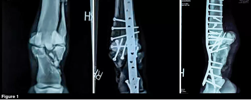
Stumbling out of the starting gate at Pimlico Race Course on May 20, he fractured three bones in and around the fetlock of his right hind leg. The very next day, he arrived at the University of Pennsylvania’s New Bolton Center where equine surgeon Dean W. Richardson reconstructed the limb. Equine veterinarian Andrew van Eps remembers the day vividly. He was working on another horse that had been brought in with a critical condition when Barbaro arrived.

“Barbaro had a terrible hind limb fracture,” he says. “The expertise, the equipment and the facilities for recovery that these horses have make it quite possible to fix complex fractures.”

The reconstructive surgery went remarkably well (Figure 1 above). By the following July, though, Barbaro had developed laminitis. The stallion underwent five more surgeries before succumbing to complications from standing limb laminitis.

When van Eps first began researching laminitis in 2000, it was widely believed that all laminitis cases were essentially the same. Today, veterinarians recognize three major forms: supporting limb, sepsis-induced and endocrinopathic. Each has a different cause and treatment protocol.

Supporting limb laminitis (SLL) is the least understood variation. A team of Colorado State University researchers published one of the only studies on the topic in 2011.1 The retrospective study included 113 horses that had casts on their limbs for different reasons.

Of those 113 horses, 12% developed supporting limb laminitis, a bit more than one in 10, van Eps says. Although the success rate for even very complex fracture repair has improved, the likelihood that SLL will develop is the main reason that treatment is not attempted in many complicated painful limb problems.

“The mortality on the development of SLL is high, at least 50%,” he says.

The Colorado State University study also indicated that the horse’s body weight and duration of casting in weeks were significantly associated with SLL.

“This is the first study to actually show that the heavier they were, the more likely they were to get it and that the longer their problem went on, the more likely it was that they would develop laminitis,” van Eps says.

Force plate studies have shown that ground reaction forces increase progressively with speed-related gait change.

Mechanical Overload Or Blood Flow?

Researchers are increasing efforts to better understand what leads to the development of supporting limb laminitis and what can be done to avoid it.

Work performed at the New Bolton Center by van Eps’ colleagues Hanna Galantino-Homer and Julie Engiles, both equine veterinarians, has shown that horses ultimately euthanized for painful limb conditions can have laminitic lesions before the development of any lameness in the supporting limb.

“I think we’re really surprised that a lot of horses have different degrees of laminitis that aren’t necessarily showing as lameness when they have a painful condition in one limb,” he says. “And it’s not always just the opposite limb, it’s often multiple limbs, which I think is really interesting.”

So the question becomes what is causing SLL to develop? Could this pathology be purely a mechanical overload problem? Or is it a blood flow problem? Could it be that they’re getting a generalized, regular laminitis-type insult that’s affecting their whole body and it’s just worse on that leg because they’re putting more weight on it? Or is it some sort of complex signaling issue?

It’s unlikely that mechanical overload on its own causes SLL, van Eps says. Force plate studies have shown that in the standing horse, the body mass is divided between the fore and hind limbs in a 60:40 ratio. The ground reaction forces increase progressively with speed-related gait change. The foot is subjected to peak forces equivalent to 0.25 body weight (bwt) at the walk, 0.5 bwt at the trot and as much as 3 bwt at the gallop.

“It is difficult to envisage a situation whereby such compensatory load redistribution can, in the standing horse, exceed the mechanical ‘strength’ of the lamellae, given that it can normally withstand up to three times the weight of the horse without adverse mechanical effect,” he says. “It’s hard to believe that we could overwhelm the mechanical properties of the tissue, but this over days or weeks sometimes, or even months, makes you feel that perhaps that could be a factor.”

Excluding mechanical overload as a significant contributing factor, van Eps points to the findings by a team of South African farriers that the interruption of blood supply under load contributes to SLL.2 The team’s study used fluoroscopy and arterial injections of contrast.

In the 1990s, Australian equine vet Chris Pollitt repeated the South African study and was able to demonstrate that when the limb and foot are under pressure, there is a decreased amount of blood flow into the capsular area. More recently, van Eps and his colleagues have also used ultrasound on the palmar digital arteries to show that the arteries can variably shut down when the opposite leg was lifted.

“They shut down at the level of the pastern, sometimes completely, sometimes partially,” van Eps says. “There’s redundancy, there are both sides which join at the bottom. Sometimes this happens on one side.”

Based on this research, van Eps focused in on the effect of weight-bearing on blood flow in the hoof using CT studies of cadaver limbs.

“First, we put some tension on the deep flexor using a spring balance,” he says, “and we looked at what was normal perfusion.”

Then the horses were loaded to the equivalent of standing square. The CT scans in this posture showed that even slightly loading the limbs was enough to interrupt blood flow.

“There was narrowing and even cut-off in the palmar digital arteries at the level just about the heel branch, but also as they enter the back of the bone to form the terminal arch in the solar canal,” van Eps says. “That gets more prominent as you increase the load so it’s equivalent to unilateral weight bearing.”

Photos: Dr. Andrew van Eps/Penn Vet New Bolton Center

Then they experimented with the effects of adding a heel wedge. In van Eps’ study, the wedge didn’t seem to affect it much. When he looked at contrast fill, this is a vascular score, so more is better. When they don’t have any load on them (Figure 2), when they have normal square-standing load (Figure 3), when they have a unilateral weight-bearing load (Figure 4), and when they have maximal load (Figure 5).

“It’s kind of quite a neat graph in terms of the score (Figure 6) and that’s the score around the dorsal aspect of the foot,” he said. “And putting a wedge, a heel wedge (Figure 7) on them, at 20-odd degrees didn’t really make much of a difference.”

Photos: Dr. Andrew van Eps/Penn Vet New Bolton Center

Blood Flow And Tissue Energy

However, he says that looking at blood perfusion is probably not enough to fully understand what leads to supporting limb laminitis.

“When we think about what the importance of blood perfusion is, it’s about supplying energy to the tissue,” van Eps says. “But then we have to study the energy balance in the live horse foot.”

Drawing from human medicine, van Eps has adapted a technique called “tissue microdialysis” to examine this relationship in the horse’s foot. In humans, this procedure is used to monitor the energy state of the brain after a traumatic injury. The method uses a tiny probe that’s roughly the same size as the smallest blood vessel in the hoof or slightly larger than a single human hair.

“We look for the tissue energy metabolites, like glucose, in the tissue,” he says. “These are brought in by the blood, lactate which is produced by a breakdown of glucose, pyruvate which goes into the oxidative side, and the ratios of those things.”

Samples can be taken as often as every 5 or 10 minutes to observe the balance of these things over time as the foot is manipulated. For example, if a tourniquet is put on a leg, the urea goes up, which means the perfusion has gone down. It’s the opposite. The glucose drops, the lactate goes up and the ratios go up (Figure 8). As another example, when the horse is walked, the urea goes down, which means the perfusion has increased; the blood flow has increased significantly.

“The interesting thing is that this does not happen in the skin,” van Eps says. “This was the skin over the tail base that we used for comparison. So it’s not just when they’re walking around that their heart rate is increased and the general blood flow is increased; this is a lamellar-specific event that’s occurring.”

Implications Of Low Blood Flow

If you measured the blood coming out of a horse’s head and compared it with the blood coming out of a foot, the foot takes more glucose per unit of blood than the brain does. This suggests that the hoof is more important than the brain to the horse, so nourishing that tissue is important.

When the foot doesn’t receive enough blood flow to nourish the laminar tissue, founder sets in quickly and dramatically on the good leg. In a recent study, van Eps and colleagues used a platform shoe that causes preferential weight bearing on the opposite limb combined with tissue microdialysis. This study showed decreased blood flow and evidence of energy starvation in the lamellar tissue, and early results support the theory that low blood flow (ischemia) plays a major role in supporting limb lameness.

LEARN MORE

Gain more insight about laminitis by:

  • Reading “Incidence of support limb laminitis in horses treated with half limb, full limb or transfixation pin casts: A retrospective study of 113 horses (2000-2009).”
  • Watching Dr. Andrew van Eps’ 2018 International Hoof-Care Summit lecture, “Understanding The Basic Mechanisms That Lead To Laminitis In Different Situations.”
  • Reading “Stopping Laminitis Cold.”

Access this content by visiting
americanfarriers.com/0918

“A combination of increased load with insufficient limb load cycling interferes with blood perfusion of the lamellae,” he says.

One unexpected finding of the study is that this is not a whole foot phenomenon. It only occurs between the lamellae. Between the lamellae toward the hoof wall, there’s less blood and more glucose being taken up by the cells. But in this experiment, there wasn’t a change in the probe that was positioned next to the bone.

“That kind of threw us off because we had a probe sitting in the sub-lamellar dermis, closer to the bone in the same feet for the whole time,” van Eps says. “And we did not see any of that occur in that zone. It was just between the lamellae.”

More Research Needed

SLL remains problematic for veterinarians treating horses with complex injuries. While the surgery to repair a fracture can have a positive outcome, the risk of limb laminitis is so significant that it prevents veterinarians from the repair.

“SLL is unpredictable, both in terms of timing and also with respect to which cases will succumb to it,” he says. “That discourages veterinarians from attempting the surgeries in the first place.”

Preventing SLL from developing is still the most effective method for treating it. And the key to avoiding it seems to be linked to the cyclic loading and unloading of feet. Future research that investigates strategies for regulating this process is likely linked to stopping it from occurring.

Such tactics could involve things like a Fitbit-like fitness tracker to monitor limb-loading cycle, improved regional pain medications, icing the feet and encouraging limb loading through walking, among others. Van Eps and his colleagues continue to study methods for improving circulation in the foot of a standing limb while simultaneously gaining a better understanding of how a loaded limb effects this flow.


References

  1. Virgin JE, Goodrich LR, Baxter GM and Rao S. Incidence of support limb laminitis in horses treated with half limb, full limb or transfixation pin casts: A retrospective study of 113 horses (2000-2009). Equine Vet J Suppl. 2011 Nov;(40):7-11.
  2. van Kraayenburg FJ, Fairall N and Littlejohn A. The effect of vertical force on blood flow in the palmar arteries of the horse. 1st International Congress on Equine Exercise Physiology, Cambridge; 1982. p. 144-54.


September/October 2018 Issue Contents