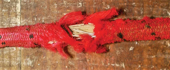
The tendons of the lower leg are one of the most crucial parts of the equine anatomy. Responsible for the elastic recall crucial for locomotion, healthy tendons are mandatory for a sound horse.

The equine limb consists of two groups of tendons, the flexors and extensors. Tendons originate from muscles above the carpus and hock; they are secured by optimally placed retinaculi as they coarse distally to their insertion. The hind superficial digital flexor tendon has medial and lateral attachment to the calcaneus of the hock. The five extensor tendons are located on the front of the forelimb, and include the digital extensor tendon, which runs along the length of the leg, while the flexors are on the backside. The flexor tendons consist of the superficial digital flexor and the deep digital flexor.

At the February 2014 International Hoof-Care Summit, Metamora, Mich., equine veterinarian Roland Thaler spoke about tendon injuries and how farriers can assist in their treatment.

“My job with this presentation isn’t so much how to shoe horses, but rather what can we do to optimize the tendon healing process,” says Thaler.

Tendon Structure

Tendons have a winded helix of structure that allows them to bear a load. They also have two distinct functions. The extensor tendons in the front of the leg are responsible for mobilization. When the muscle contracts, the tendons protract.

“The extensor tendons provide little support function,” says Thaler.

“On the other hand, flexor tendons located in the back of the leg, such as the super digital flexor tendon, are primarily supportive.”

The deep digital flexor tendon is capable of more mobilization than the other flexor tendons. Its movement causes retraction, which propels a horse forward. It also provides support to the fetlock.

The primary function of a tendon is to transfer high tensile loads, but Thaler says they also have other functions. For example, tendons can take a group of different muscles and link them together to form a common unit. Tendons can also take energy and disperse it across several different bones.

Tendons are made up of 50% to 60% water, tendon cells, collagen, extracellular matrix and blood supply. Tendon cells start as tenoblasts, and as they mature, they become less metabolically active. This enables them to sustain a large energy load without using a lot of oxygen.

The strength of a tendon is based on its roundness and size. Strength is also dependent on collagen formation because healthy collagen forms the major structural units of the tendon. Environmental effects also determine strength. If a horse exercises hard, the tendons don’t change much in size, but they do change in composition.

footing.jpg

Choosing the appropriate footing is critical for when the horse is rehabbing a tendon injury.


With exercise, collagen turns over. The tendon will change its physiology, but not its structure so much.

“But we do know that if you stop exercising the horse, if you put that horse in a stall, if you put a cast on its leg, that extracellular matrix degrades very quickly and it just takes a few weeks and it’ll take months to get that back,” he warns.

Without use, the tendon becomes stiffer due to increased production of degenerative enzymes and decreased blood flow. Therefore, when it comes to tendon injuries, rest causes more harm than good.

In human medicine, the practice of treating a sprained ankle with a cast and crutches is outdated because doctors have discovered that it’s important to keep tendons and ligaments loaded and moving during the healing stage. The main function of exercise in treating tendon injuries is to increase circulation, which in turn stimulates healing.

Causes Of Injury

Tendons get damaged in three different ways: direct trauma, such as a wire cut; tensile overload, which is excessive strain of the tendon structure during a workout or race; and complex, which is progressive trauma to a degenerative structure, such as the chronic active tendonitis of the deep digital flexor tendon of a show jumper.

Trauma often happens when a horse kicks through a fence and cuts through a tendon, or kicks a board in a trailer. A horse also injures itself through tensile overload while running across a field and hitting a hole, or stepping sideways and tearing a structure.

Typically, though, tendon damage is a complex issue.

“There’s degeneration in that tendon, and then there’s some kind of activation or acuteness,” says Thaler. “So we get this hot tendonitis and we have a hot acute injury, but it’s the result of this acute injury put on top of a diseased tendon.”

Tendons are strong, and are good at withstanding muscular contraction and muscular physiologic loads. When an injury occurs on a normal tendon, the area of damage is often at the muscle tendinous junction, or at the insertion of the bone. Most of the injuries vets see are in the middle area of the tendon, which means the tendon wasn’t healthy before the injury. These tendons are often diseased, have chronic inflammation and are connected to joints with degenerative arthritis.

“So you get this kind of disease of the tendons, it’s compromised and the horse works a little bit too hard for the compromised tendon and then it becomes injured,” says Thaler.

If a horse has a diseased tendon and then suffers an injury, the horse has more than an 80% chance of re-injury.

“So no matter what you do, that tendon is going to probably get hurt again. It has a diminished capacity to heal,” he says.

Issues with the tendon can have a number of causes, but most are mechanical. An example is chronic overloading, where the horse is working hard and experiencing too much tension. Tendons also can break down for physiologic reasons, like Cushing’s disease.

Fixing Tendons

Tendon repair is controlled by the tendon cells and by the extracellular matrix, which is the same healing process as with most connective tissues. A scar often results with this type of healing. Healing comes in three phases: inflammatory, proliferative and remodeling.

The inflammatory phase typically lasts about 5 to 7 days, and this is when the tendon is at its weakest point.

“Say you’re at a horse show and that tendon is starting to get hot and sore, and it has just that one more class. The trainer wants to know if we can get it through that class — no. But a lot of people push that and I think a lot of trainers know the tendon is getting a little hot, a little sore and they kind of make that judgment call to push to get through this class, hoping that they can then have all the time off the horse needs. It’s a bad decision because the tendon is at its weakest.”

During the inflammatory phase, inflammatory cells, blood cells and fibrin debris take part in the process. A hemotoma may also form at the sight of the injury. The tenocytes become activated and produce collagen 3, which matures and eventually becomes stronger.

During the proliferative stage, cells start rebuilding. The tendon cells begin creating both collagen and extracellular matrix. Proliferation comes from the blood supply and forms a large knot in the fibrous tissue called a tendon callus. This callus creates the classic appearance of a bowed tendon.

The proliferative phase lasts from 1 to 6 weeks and starts the first week after the injury. At 2 weeks, the tendon has usually developed enough callus to start healing.

At 3 to 4 weeks, the limen of the scar starts, and once there is stability, it starts shrinking back and will continue to do so for the next 20 weeks. When a section of healing tendon is examined under a microscope, it resembles a normal tendon and will have a similar structure.

“But physiologically, a normal tendon and a healing tendon are very different,” explains Thaler. “The physiologic change caused by the damage means that tendon will never likely return to its original strength.”

Diagnosis

The most common way to diagnose tendon injuries is through clinical examination. Heat, pain and swelling are signs of an injury, and are the result of the tendon protecting itself from pressure and tensile overload.

ultrasound

Thaler finds ultrasound is an effective tool in treating tendon injuries with periodic usage.

Although clinical examination is important, it does not reveal how the age of the injury or how the disease is progressing. Thermography is a good tool for making these determinations, but is difficult to use in the field.

Magnetic Resonance Imaging (MRI) also is a good method for diagnosing tendon injury, but is expensive and impractical for many horse owners. Consequently, the most commonly used method of tendon problem diagnosis is ultrasound.

Ultrasound reveals fluid content, enlargement of the structure, and change in architecture.

“The advantages of ultrasound are that it’s very sensitive, very specific and many veterinarians have access to it,” says Thaler. “The disadvantage is that the results can be subjective. The quality of the ultrasound is dependent on the operator.”

However, ultrasound as a diagnostic tool is improving, in part because equipment is improving. Training in the use of ultrasound is also getting better, and techniques are refining.

A new technology called Ultrasound Tissue Characterization (UTC) was designed by Hans Van Schie, a professor at Monash University in Australia and director of research at UTC Imaging in the Netherlands.

“UTC is being used to evaluate the Dutch Olympic team horses once a month,” says Thaler. “This type of ultrasound machine has a track that goes against the back of the leg while the ultrasound scans down for 40 seconds, and then transfers the results into a three-dimensional image.”

It measures fibril length, and characterizes the quality of the tendon. If a horse is working hard, a little bit of edema will show up in the tendon, and the horse can be given 2 or 3 days of rest along with ice. The UTC can also show whether the tendon is starting to degenerate. UTC has just become available to veterinarians in the future.

Treatment

Treatment of tendon injuries is a delicate balance of stability and mobility. If the horse is asked to do too much too soon, the tendon is re-damaged. Too much motion causes a decrease in healing. If the horse doesn’t move enough, the tendon won’t heal. At 7 days post-injury, if the vet instructs the horse owner to walk the horse and the tendon is disrupted, healing is delayed.

The key to treatment is to specifically identify the lesion through MRI or ultrasound. Knowing which structure it is, the severity of the injury and the age of the injury are crucial.

It’s important to manage the inflammatory stage in the first week after the injury. Cold therapy is best, preferably cooling the leg at least 10 minutes at a time, preferably 60 minutes four times a day. Ice boots are ideal for this, while gel packs are less effective. A minimum of a 10-degree difference in Celsius temperature is needed for the therapy to be cold enough. In other words, if it’s 60 degrees outside, running a hose on the leg will not provide enough cold. Cold applied with pressure is best because by putting pressure on the area, the venous return is decreased and the blood flow is slowed, helping to cool the area more effectively.

Injured tendon protocol used to include anti-inflammatory drugs, but human tendon surgeons found that non-steroidal anti-inflammatories decrease the quality of healing. It’s been demonstrated that a 20% decrease in strength occurs when humans receive anti-inflammatories after tendon surgery, and incidence of complications is 40% greater.

Biomechanical optimization is an option for treatment and involves farrier work. The farrier’s goal is to modify the loading on the tendon to facilitate healing by modifying loading during the healing process. Different farriers have different ideas on how to do this. Most base it on the horse’s comfort.

Biomechanical optimization can change the foot simply through trimming. Appliances can also be added to the foot, such as wedges. “Asymmetric shoes can also help, along with having the horse walk on soft footing,” says Thaler. “For a horse with a deep flexor tendon injury, a reverse shoe may be called for. As the horse is beginning to heal, an onion shoe can help. In time, a sport shoe may work best.”

It’s important to remember that increasing the palmar angle will reduce the tension of the deep flexor-associated structures. This includes the inferior check ligament and navicular structures. Conversely, by decreasing the palmar angle, tension is reduced in the suspensory ligament and superficial digital flexor tendon.

Rehabilitation is the next step in treatment. The first week after the injury, hand walking is important. Horses with swelling or sensitivity should be hand walked right away. (In horses that are too uncomfortable to walk, stall rest is warranted.)

During week 2 to week 6, hand walking should be the only exercise, and only on a surface that assists in the healing. If the horse has a deep flexor tendon injury, it should be walked on softer footing. In the case of a
superficial flexor tendon or a suspensory ligament injury, the horse should be walked on firm footing. Horses fitted with asymmetric shoes, an onion shoe, a reverse shoe or an egg-bar type shoe should be walked in soft footing.

An ultrasound at 6 weeks reveals whether the tendon has stopped degrading, and whether the inflammation is coming down. An ultrasound can indicate whether the defect field has filled up. Determining when to start the horse back to work or change the shoeing will depend on whether this defect has filled. The horse should then be ultra-sounded every 6 weeks to determine if there is fiber alignment. Once this becomes evident, the farrier can return to standard shoeing, and the horse’s workload can be increased.

By 20 weeks, most of the tendon tissue should appear healed under ultrasound. The healed tissue is not as strong as original tissue, so even though the tissue is healed ultrasound graphically, the horse is probably not ready to go back to its original job. The horse either requires more time and rehabilitation, or a lighter workload.

 “I think if we can recognize that tendon injuries are degenerative in nature and we can approach it that way, and know that something repaired is not as good as brand new, then we can more accurately help our clients attain what they’re trying to achieve,” says Thaler. “They just want to see this horse perform again, and we just need to show them how.” 


Learn More

Why Equine Bones Break and Tendons Rupture

Understanding anatomy and physics can help prevent severe injuries.