Imaging technique can provide valuable information prior to P3 displacement
Terms For Measurements On Lateral Radiographs: Distance In Millimeters (mm)
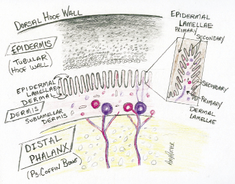
Epidermis – Horn of the hoof capsule (or skin).
Dermis – Soft tissue: contains blood vessels, nerve, lymphatic vessels and connective tissue.
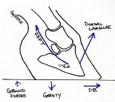
Dorsal – Toward the front of the hoof capsule (ex: dorsal hoof wall).
Palmar – Toward the back or bottom of the hoof (ex: heel or sole).
Medial – Toward the midline (“inside”).
Lateral – Toward the “outside.”
P3 – Coffin bone/third phalanx/distal phalanx.
DDFT – Deep digital flexor tendon.
Proximal – Closer to the body.
Distal – Away from the body (toward the ground).
Horn-Lamellar Zone (HL): Width of the dorsal hoof wall when measured perpendicular to the face of P3. Half the width is horn (epidermis — diseased in white-line cases) and half is lamellae (dermis, which contains the blood vessels — diseased in laminitis.) A healthy 2-year-old Thoroughbred foot has a HL measuring 15 mm at the proximal and distal aspect of the face of P3. This number will be larger with certain breeds (warmbloods, draft horses), or increases with disease.
Coronary-Extensor Distance (CE): Vertical distance from the level of the coronary band to the top of the extensor process of P3. If a horse is a “sinker,” this number may increase in a matter of days as P3 descends in the hoof capsule. The CE may also slowly increase in cases of chronic laminitis due to collapse of the P3 suspension and resultant capsule distortion.
Laminitis destroys the attachment between P3 and the hoof capsule. The forces of the deep digital flexor tendon continue to pull the apex of P3 away from the dorsal wall and down onto the solar dermis. This compresses the blood supply of the sole, reducing sole growth. Further collapse of the foot results in alterations in wall growth, bone disease, abscesses/seromas, etc.
Sole Depth (SD): Depth of sole measured directly below the apex/tip of P3. If the foot is strong, I’ll include the measurement of SD + cup. On radiographs the “cup” of the foot appears as a black space distal to the sole. I encourage sole growth: the heavier the sole, the greater protection for P3. I also correlate increases in SD with health. (If the load is controlled within a foot, and the dermis has a good blood supply, it will grow sole.) There is a wide range of “normal” in SD, but while a thin sole may be common, it is not healthy. Although many horses race with 10 mm SD, that foot isn’t optimal. In a foot with a heavy sole, SD will approach 20 mm. A foot with a heavy sole will have a deep sulcus around the frog (even at the apex) and usually has a cup and a strong wall.
Example Horse: 14-year-old warmblood gelding. Chronic lameness due to rim fractures on distal border of P3 when adopted 7 years ago. Poor wall quality, difficult to maintain sole depth and mild arthritis. Rotational limb deformity (toes out) and crushed medial heel.
CE: Coronary-Extensor Distance –13 mm.
HL: Horn-Lamellar Zone – 19-20 mm.
SD: Sole Depth – 17 mm.
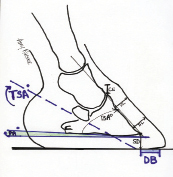
DB: Digital Breakover – 23 mm.
BA: Bone Angle – 45 degrees.
PA: Palmar Angle (measured to low medial wing of P3) – 3 degrees.
TSA: Tendon Surface Angle – 30 degrees.
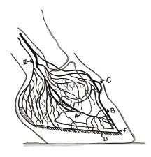
A: Terminal arch of palmar digital vessels.
B: SLVB - Sublamellar vascular bed.
C: Coronary plexus.
D: Circumflex vessels (with sole papillae).
E: Heel vessels.
F: LCJ – Lamellar/Circumflex junction.
*Digital Breakover (DB): Distance measured by dropping a line from the apex of P3 to the ground surface (DB=0) and then measuring anterior to the front of the toe (the place the foot is last in contact with the ground as it moves forward). DB is manipulated by trimming the foot (rocker toe) or shoeing techniques (rocker or bevel toe, rocker shoe, etc.). DB may be negative if the breakover is behind the apex of P3. We manipulate/reduce DB to reduce the lever-arm forces of the toe on the dorsal wall and lamellae, as well as the joints, DDFT, and navicular apparatus.
*Palmar Angle (PA): Angle between the palmar surface of P3 and the ground. The PA may be positive (for example, a clubfoot), zero (palmar surface of P3 is parallel to the ground) or negative (wings of P3 are “lower” than the apex in a low-heel, long-toe foot). Note: Increasing the PA will decrease the strain of the DDFT, reducing the pull of P3 away from the dorsal wall and down onto the sole dermis.
Venogram: Folded lamellar-circumflex junction, loss of sole papillae.


