“Horse Conformation: Principles of Form and Function” (2022), represents a significant expansion and upgrade of my “Principles of Conformation Analysis” (1986). It’s a large-format, full-color, three-volume set containing over 900 pages of information. There are hundreds of photos of horses with good vs. poor conformation plus charts, diagrams, analyses and detailed dissections that clarify the link between anatomy on the inside and conformation visible on the outside.
Volume I explains basic anatomy and function and includes growth and maturation and how to predict what a foal will look like when it is mature. Volume II looks at the back, loins, withers, ribcage, neck, skull and teeth and includes a chapter on saddle fitting. Volume III focuses in detail on the fore and hind limbs and hooves. Each volume presents a “twinned” structure — first there is an overview chapter that explains how a particular body part works, then this is followed by a chapter showing good and bad conformation.
The following is an excerpt from Chapter 16 in Volume III, which pertains to the equine forelimb. In the book, this chapter covers a huge amount of material, including everything that can go “wrong” from the withers to the coffin joint — a virtual catalog including steep shoulders, mutton withers, sweeney, capped elbow, bench knees, knock knees, bowlegs, small round joints, big knee, boggy knees, calf knee, bucked vs. buckled knees, splints, bucked shins, suspensory tear, osselets, upright pasterns, splayfoot, windpuffs, ringbone, sidebone and pathologies of the navicular sesamoid. In this review, a fraction of the total package is presented, all of which are of interest and practical use to the farrier. Order Dr. Bennett’s books by emailing office@equinestudies.org.
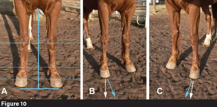
Pastern conformation is to be evaluated in the same way as the conformation of the carpus, that is, by first visualizing the plane of assessment, and then by scanning for rotations and deviations (offsets are rarely found at this joint or the coffin joint). The pastern segment articulates downward with the coffin bone. First check at the level of the fetlock joint, then at the level of the coronet band. Tilting at either level indicates medial or lateral deviation, while rotations can be picked up by visualizing the plane of assessment. Rotation at the level of the coronet band is accompanied by a spiraling or swirling pattern to the hair.
Offset or “bench” knees in a warmblood. The top of the cannon bone is not centered beneath the carpus.
A rear view of the pasterns and heel bulbs often reveals more than the front view. The center of the fetlock joint should be positioned directly above a point midway between the heel bulbs, and not off to the medial or lateral side. If the hairline that runs down the center of the back of the pasterns leans toward the inside, or if the hairs have a spiraling orientation, they signal misalignment. Scan also for the heel bulbs’ height; differences in height signal deviation in pastern angle and also problems in the interior of the hoof capsule (i.e. sheared feet).
Pastern length in domestic horses varies from about 10% to about 20% of the body length, with the vast majority falling from 13-17%. Because equine pasterns are relatively long, they exert considerable leverage during locomotion. The muscles that support the fetlock joint and move the pastern segment are the SDF and DDF, whose tendons run over the slick, smooth, saddle-shaped intersesamoidean “ligament” that lies between the sesamoids at the back of the fetlock joint. It is primarily the tonus or “spring back” quality of the flexor muscles that keeps the fetlock joint from descending to the ground, assisted by the suspensory apparatus. Fatigue of the flexor muscles is the precursor to hyperextension of the fetlock joint and thus to overstretching and tearing of the suspensory yellow ligaments. The resting angle of the pastern ranges from as steep as 70 degrees to horizontal, with most horses measuring between 47 and 60 degrees. Horses with “soft pasterns” usually also have coon-footed conformation, which means the resting pastern angle is lower than 40 degrees. The pasterns, either “soft” or coon-footed, may or may not be longer than average; they are never desirable.
Pasterns that are literally upright, i.e. steeper than 70 degrees, almost certainly reflect either positive or negative rotation of the coffin bone and concomitant misalignment of all the bones above; in other words, they are pathologies, not part of the animal’s native conformation. “Short, upright pasterns” are fingered as the culprit in a plethora of pathologies and movement problems, again almost always wrongly. The buyer need not be afraid of pasterns in the steeper part of the normal range, so long as the fetlocks are normally flexible, the horse is serviceably sound, and the distal limb is free of pathology as.
Navicular syndrome is especially problematic. The terms “navicular syndrome” or even just “heel pain syndrome” are now being used instead of “navicular disease” because X-ray diagnosis of lesions specifically affecting the navicular sesamoid can be difficult, and because other types of scans (scintigraphy, MRI and CT) have shown that pain in the rear portion of the horse’s foot may or may not be caused by or associated with degeneration of the navicular sesamoid.
Upright pasterns are often cited as predisposing to “navicular,” but horses with steep pasterns are no more likely to be affected by navicular syndrome than those with any other normal pastern conformation. Massiveness, or a high ratio of body weight to hoof width such as often seen in both warmbloods and Quarter Horses (see Chapter 4), is however a significant contributing factor. So are flat feet and weak or thin hoof walls, which occur in many Thoroughbreds. Horses with thick, strong hoof walls that have been suffering from heel-pain syndrome for some time may develop thin digital cushions while the hoof becomes boxy and upright. Chronic unweighting of the heels leads to contraction of the heels, which adds to the compression and pain already present in the caudal part of the foot.
When heel-pain syndrome involves degenerative change to the navicular sesamoid, a primary cause is compression of the navicular sesamoid and coffin joint by the DDFT. Constant friction erodes first the protective cartilage and then the sesamoid itself. Inflammation and degenerative changes to the navicular sesamoid produce characteristic lollipop-shaped deficits within the bone, while the surface of the bone facing the DDFT becomes rough and prickly, causing intense pain. When both front feet are affected about equally, the horse often adopts a characteristic “sawhorse” stance with hindlimbs angled back, forelimbs angled forward. When it is more severe in one foot than the other, the animal will stand with the more painful foot placed ahead of the other, a stance which is termed “pointing.” It may deliberately seek soft mud or other soft footing to stand in. The most revealing posture is when the horse tries to take weight off its heels by “knuckling forward,” as if it were trying to pull its limb out of its hoof capsule (as a person would take their foot out of a slipper).
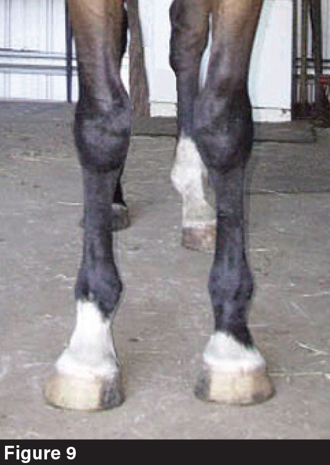
Developing a better-informed standard of horse show judging would create a sea change in all of this. I am speaking of something that would go beyond merely improving judging technique as shown in Figures 9 and 10. A proper standard would prize horses with feet of the correct size and normal shape and punish those with inadequate substance compared to bodyweight, limb substance that diminishes downward, small feet and weak-walled hoof capsules. Such horses should not be bred, even if they are spectacularly muscled, devastatingly pretty, or happen to be the fastest thing on the track. I do not expect this advice to be relished by movers and shakers in horse breeding; but until it is taken seriously, we will continue to have wastage, the cost of which is ultimately absorbed not only by the suffering animals, but by ordinary horse owners with ordinary incomes.
Read more from Dr. Deb Bennett in "Conformation Analysis of the Equine Forelimb" in the November 2025 issue of American Farriers Journal.


