
American Farriers Journal
American Farriers Journal is the “hands-on” magazine for professional farriers, equine veterinarians and horse care product and service buyers.
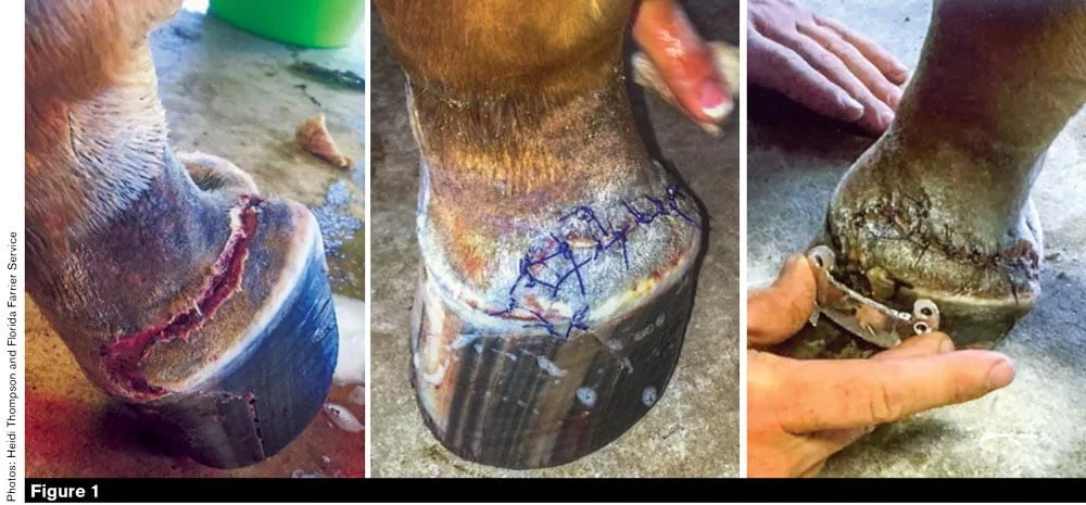
Heidi Thompsons’ 18-month-old filly Maybeline lacerated her front left foot on a barb-wire fence in early June 2017. Veterinarian Dr. Valerie Biehl examined the horse, sutured and treated the wound.
This case involves my client Heidi Thompson’s 18-month-old filly, Maybeline. On June 9, 2017, she was turned out after it had been raining. She was out in the arena because it offered the driest surface at that time. Maybeline managed to find a puddle and slip. Her foot went under a wire fence, which lacerated the medial side of her hoof. Dr. Valerie Biehl, Maybeline’s veterinarian, came out to examine and suture the wound that day.
When Heidi contacted me to come by about a week later, the stitch pulled and Maybeline ended up with quittor in the medial wing of her coffin bone. At that point, I advised Heidi that the animal most likely needed to go to surgery. Heidi did not want to pursue surgery, so my next thought was that Maybeline should probably be euthanized.
“Clayton, that’s not an option,” Heidi told me. “Fix her.”
My main objective in a…

American Farriers Journal is the “hands-on” magazine for professional farriers, equine veterinarians and horse care product and service buyers.

American Farriers Journal is the “hands-on” magazine for professional farriers, equine veterinarians and horse care product and service buyers.
Download these helpful knowledge building tools

We are here to support you.
We stock a wide range of high-quality products from trusted brands to ensure durability, performance, and reliability in every job you undertake. Our extensive inventory of horseshoe products and farrier tools means you can find everything you need in one place, saving you time and effort. Your satisfaction is our top priority. We are committed to providing excellent customer service, prompt shipping, and hassle-free returns.