American Farriers Journal
American Farriers Journal is the “hands-on” magazine for professional farriers, equine veterinarians and horse care product and service buyers.

Venograms aren’t the most commonly used type of imaging in hoof care, but there are veterinarians who would like to see them used more often.
Proponents of venograms are using them not just as a diagnostic tool, but also to help guide the course of care. Others believe venograms could offer intriguing research possibilities.
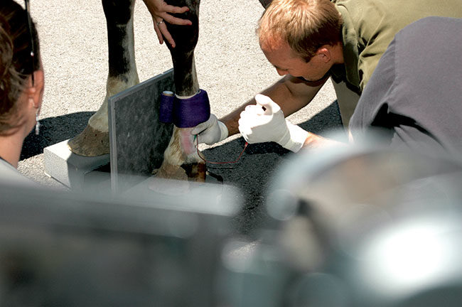
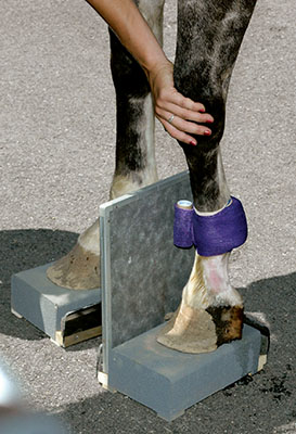
Venograms involve taking an X-ray of a particular part of the body after a dye has been injected into a vein. In hoof care, venograms are typically done in the lower limb and foot. The dye, which provides a contrast in the radiograph, allows the veterinarian to view the blood supply in the hoof.
Venograms have been used most commonly as part of diagnosing and treating laminitis.
Christopher Pollitt, a veterinarian and researcher from the University of Queensland in Australia and Ric Redden, an equine veterinarian from Versailles, Ky., pioneered the procedure in standing horses in 1992.
The biggest drawback with venograms may be learning to read them. Redden, an advocate of their use, says reading venograms is a skill that can only be mastered through experience and repetition.

A radiographic plate is already in place before the dye is injected, because the X-rays need…