Hoof abscesses are probably the most common cause of acute lameness in horses encountered by veterinarians and farriers.
A hoof abscess can be defined as a localized accumulation of purulent exudates located between the germinal and keratinized layers of the epithelium, most commonly subsolar or submural.
Much debate still abounds between the veterinary and farrier professions as to who should treat a hoof abscess and the best method in which to resolve the abscess.
Remember the origin of the organisms that are responsible for a hoof abscess gain entry through the hoof capsule (epidermis) into the inner subsolar/submural tissue (dermis) where the organisms initiate an abscess. Foreign matter (such as gravel, dirt, sand and manure, coupled with infectious agents such as bacteria or fungal elements) generally gains entry into the hoof in one of three ways:
- Through a break or fissure in the sole-wall junction (white line).
- A misplaced nail or a puncture wound somewhere in the solar surface of the foot.
- By way of a full thickness hoof wall crack or multiple old nail holes.
Formation Of An Abscess
It may be easier for us to understand how to treat an abscess if we briefly look at the mechanism by which an abscess forms.
Foreign debris will gain entry and accumulate in a small separation or fissure
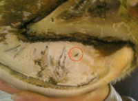 Abscesses form when foreign debris gain entry and accumulate in a small separation or fissure located in the sole-wall junction anywhere around the perimeter of the foot.
located in the sole-wall junction anywhere around the perimeter of the foot, including the inner surface of the bars adjacent to the sole (Figure 1).
As the animal bears weight, foreign matter will migrate through the fissure until it reaches the subsolar or submural tissue (dermis). Once inside the hoof capsule, the defense mechanism within the dermal tissue recognizes the matter as foreign and sets off a reaction. The bacterium contained within the debris invades the dermal tissue and leads to inflammation, the bacteria continue to grow and cause neutrophils (white cells) to migrate into the area.
Enzymes released from the bacteria and from the invading white cells lead to liquefaction tissue necrosis and the development of the gray/black exudate. The inflamed area is quickly walled off with a thin layer of fibrous tissue to form an abscess. The inflammation and the pressure from the accumulation of the exudate exerted on the surrounding tissue leads to the clinical signs associated with a hoof abscess.
Dermal tissue can be inoculated by bacteria from a misplaced nail in two ways. The nail can be driven directly into the laminar corium. When the nail enters dermal tissue, the horse will show discomfort as the nail is driven into the foot and there will be hemorrhage present where the nail exits the outer hoof wall.
Blood observed at the exit of the offending nail will alert the farrier of the misplaced nail and the blood also acts as a "physiologic rinse" to dilute or eliminate bacterial contamination. Removal of the nail and application of an appropriate antiseptic will usually prevent infection.
Another scenario that occurs frequently is that while the farrier is driving a nail, the horse shows pain indicating the nail is invading sensitive tissue. The farrier will generally remove the nail, place it in another spot or direction and again drive it into the foot. When this occurs, the farrier should remove the shoe and examine the spot where the nail entered the foot. When a nail enters dermal tissue (even if removed), it can seed the area with organisms and lead to an abscess.
If the nail has entered the foot inside the sole-wall junction, the owner should be alerted as to potential problems and the horse could be placed on a broad-spectrum antibiotic for 3 to 5 days as a prophylactic measure.
Finally, we have the condition described as a "close nail" where the nail is placed so that it lies against the border of the dermal corium just inside the hoof wall. Pressure against the corium, the movement of the horse combined with the organisms introduced with the nail, will lead to an abscess as described above. There is a lag period of 7 to 14 days or even longer before clinical symptoms or discomfort is observed following the placement of a "close nail."
Another common cause of perceived subsolar abscesses is penetration of the bottom of the foot (sole) by a sharp object. This is not actually an abscess but rather a diffuse infection caused by the solar corium being seeded with organisms from the penetrating object.
Pain is immediate and usually followed by infection within 3 days. A full-thickness puncture wound in the sole always requires veterinary input.
Clinical Signs
Most affected horses show sudden (acute) lameness. The degree of lameness varies from being subtle in the early stages to non-weight bearing. The digital pulse felt at the level of the fetlock is usually bounding and the involved foot will be warmer than the opposite foot. With careful observation — unless the abscess is in the middle of the toe — the intensity of the digital pulse will be much stronger on the side of the foot where the abscess is located.
If the abscess is long standing, there may be soft tissue swelling in the pastern or above the fetlock on the side of the limb corresponding to the side of the foot where the abscess is located.
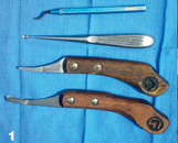 Drainage of an abscess is accomplished by opening the offending tract or fissure using a thin small loop knife, a 2 mm bone curette or other suitable probe.
The site of pain can be localized to a small focal area through the careful use of hoof testers. Sometimes with acute lameness, the pain will be noted over the entire foot with hoof testers and, in this case, veterinary assistance is used to rule out laminitis, a severe bruise or even a possible fracture of the distal phalanx (P3).
Treatment
The most important aspect of treating a subsolar/submural abscess is to establish drainage. The opening should be of sufficient size to allow drainage, but not so extensive as to create further damage.
When pain is localized with hoof testers, a small tract or fissure will commonly be found in the sole wall junction (white line). The wound or point of entry may not always be visible, as some areas of the foot such as the white line are somewhat elastic and wounds in this area tend to close. In this case, a suitable poultice should be applied to the foot daily in an attempt to soften the affected area and eventually a tract will become obvious.
The offending tract or fissure is followed within the white line using a thin small loop knife, a 2 mm bone curette or other suitable probe (Figure 2). The tract is slowly followed until a gray/black exudate (pus) is released and the probe will enter the "belly" of the abscess. At this point, the tract is open into the cavity of the abscess.
A small opening is all that is necessary to obtain proper drainage (Figure 3). This can be determined by placing thumb pressure on the solar side of the tract and observing more drainage being expressed or a bubble at the opening when pressure is applied. Care should be taken to avoid exposing any corium, as it will invariably prolapse through the opening, prevent closure of the tract and create an ongoing source of pain. Under no circumstances should an abscess be approached through the sole.
The draining tract can be kept soft and drainage promoted in many ways. The application of an Animalintex poultice that has been soaked in hot water is applied for the first 24 to 48 hours has been useful in the author's hands. This is a self-contained, medicated poultice, which is commercially available through your veterinarian or tack shop.
The author prefers the sheet version of this poultice rather than the poultice pad distributed by this company, as the whole foot, including the coronet, should be enveloped in the poultice.
Using Soak Bandages
Another method to encourage drainage is to apply a soak bandage. Here layers of practical cotton are crisscrossed to form a heavy bandage that envelops the foot. MgSo4 (Epson salts) is placed in the inner foot surface of the bandage and the bandage is attached to the foot.
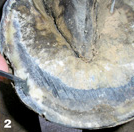 A small opening created to establish drainage. Note area of hoof wall separation (so-called white line disease) palmar to the draining tract.
The bandage is now saturated with hot water and saturated periodically over the next 24 to 48 hours. Using either of these methods eliminates the need for continued foot soaking.
Ichthammol ointment is a coal tar derivative with mild antiseptic properties that has been described for treating skin disease in both humans and animals. The use of an Ichthammol bandage for treating hoof abscesses, both before and after drainage, has become another traditional treatment among veterinarians and horse owners with reportedly good results. The author has not used this form of treatment, therefore would be unable to render an opinion as to its efficacy.
The tetanus immunization status of the horse should always be determined.
The horse should show marked improvement within 24 hours. Following the poultice or foot soak bandage, the hoof is kept bandaged with an appropriate antiseptic such as Betadine solution/ointment or 2 percent iodine until all drainage has ceased and the wound is dry. At this point, the opening is filled with Keratex Hoof Putty that keeps the affected area clean and prevents the accumulation of debris within the wound. The shoe is replaced when the horse is sound.
If The Infection Migrates
Many times the painful tract can be located, but drainage cannot be established at the sole-wall junction. In this case, the infection is deep and may have migrated under the sole or wall away from the white line. Again, under no circumstances should an opening be created in the adjacent sole. This seldom leads to the abscess, generally leads to hemorrhage and may create a persistent, non-healing wound with increased potential for bone infection.
Instead, a small channel can be created on the hoof wall side of the white line using a small pair of half-round nippers. The channel is made in a vertical direction following the tract to the point where it courses inward. Drainage can usually be established using a small probe in a horizontal plane. Preferably this is done at the onset of lameness, before the infection ruptures at the coronet.
If left untreated, a hoof abscess will follow the path of least resistance along the outer margin of the dermal tissue and eventually rupture at the coronet forming a draining tract. Many horse owners actually consider this to be an acceptable practice and wait for this to take place. From a humane standpoint, this practice often extends the amount of time the animal experiences severe pain.
Rupture at the coronet also leads to a permanent scar under the hoof wall. This tract leading to the coronet may result in a prolonged recovery from the abscess, a chronic draining tract, repeated abscesses and a full-thickness, hoof-wall crack. Every effort should be made to establish drainage on the solar surface of the foot.
Abscess Or Infection?
Please be advised that the following comments are the author's opinion and do not reflect the position of the American Association of Equine Practitioners (AAEP) or any other veterinary medical organization.
Members of the veterinary and farrier professions have debated the topic of who should treat hoof abscesses for ages. If we go back and consider how an abscess is formed, it is a cavity filled with exudate surrounded by a thin fibrous membrane.
The cavity of the abscess could be thought of as an extension of the entry tract located in the hoof capsule. Therefore, when a farrier follows a tract through the sole-wall junction and creates a small opening into the cavity of the abscess, he or she may not be invading dermal tissue.
There is no hemorrhage or pain involved with this process. It could be considered much the same as removing a splinter from under the skin in a person. In this context it would appear justified for a farrier to drain an abscess and initiate the aftercare described previously.
Again, it could be argued and/ debated whether this is the practice of veterinary medicine. Furthermore, it would be prudent and in the farrier's best interest to inform the horse owner at the onset as to his or her intention of draining the abscess, to give the owner the option of contacting a veterinarian and explaining to the owner that hoof abscesess can and often do persist to a point where veterinary intervention would be necessary.
On the other hand, when an infection is present from a puncture wound in the sole or a "close nail," the treatment should be a joint venture with a veterinarian.
To establish drainage in this case, a larger opening may need to be created and sedation may be necessary, dermal tissue will need to be invaded and possibly debrided, there may be hemorrhage and medications such as antibiotics and anti-inflammatory drugs will need to be prescribed.
If a farrier were to treat an established infection in the hoof, it would be practicing veterinary medicine and the farrier could be held liable.
Prevention
Prevention is achieved through proper hoof care and centers on promoting a strong, solid sole-wall junction (white line) that resists penetration by debris. Hoof abscesses are less likely to occur when a solid sole-wall junction (white line) is maintained.
Excessive toe length increases the bending force exerted on the toe, leading to a widening and weakening of the white line. Other conditions that cause mechanical breaks or weakness in the continuity of the white line are hoof capsule distortions (long toe-under run heels, excessive toe length, heels too high or a club foot, sheared heels), hoof wall separations (white line disease, seedy toe) and chronic laminitis. Excessive moisture or dryness may also contribute to weakness in the white line.
Proper Trimming
To prevent abscesses, it is important that the foot be trimmed in a manner that accentuates a strong healthy foot. A few basic principles can be used when trimming to create a strong foot and strengthen the white line.
First is the creation of a good heel base where the bars are preserved and the heels are trimmed to the base of the frog, or as far back as possible. This increase in ground surface allows a substantial amount of weight bearing to occur in the palmar portion of the foot. Sole is only removed adjacent to the white line to identify excess hoof wall that should be removed. It is not necessary to concave the sole as this occurs naturally.
The toe is trimmed appropriately and backed up from the dorsal surface (front) of the hoof wall, such that a line drawn across the widest part of the foot will be in the middle of the foot.
This assures that there is no excessive toe length. In some cases, fitting the shoes hot may be helpful to seal the sole wall junction. The use of hoof hardeners (Keratex) and bedding the horse on shavings or sawdust may be useful to harden the feet during extremely wet weather or when the horse is being washed frequently, such as during horse shows.
During dry weather, a hoof dressing, such as a combination of cod liver oil and pine tar (mixed in a ratio of 3:1) painted on the entire foot, may help to soften the hoof capsule.
Preventing indirect penetration through the white line is therefore dependent on providing adequate protection to the underlying sensitive structures. The hoof capsule has a natural ability to provide such protection and it is imperative that we strive to enhance these strong features through proper trimming. Excessive removal of protective horn is a common practice, as emphasis is often placed on eye appeal instead of functional strength.


