Takeaways
- Successful treatment of severe hoof conditions like osteomyelitis and sepsis relies heavily on a close, coordinated partnership between veterinarians and farriers.
- Advanced farriery using heart-bars, impression material and casts are crucial for redistributing load-bearing and protecting compromised structures.
- Recovering from extensive equine hoof wall resections requires patience, diligent medical management and long-term supportive care.
Around mid-August 2021 and needing a well-earned 3-day break to the South African bush, the phone rings. On the other end of the line is Dr. Andrew Gray, director and chief veterinary surgeon at VetScape Referrals just outside of Cape Town, South Africa. He’s calling me because there’s a hoof-related emergency at the hospital.
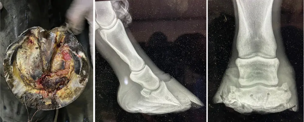
After a couple more phone calls, farrier Alan Theron, CPF, arrives, and a shoeing plan is formulated over the phone for a well-bred Thoroughbred mare that is heavily pregnant to a top stallion. Her near fore is a total septic mess with a massive sub-solar abscess and already evident necrotic tissue (Figure 1).
Gray surgically removes a large necrotic sequestrum from P3 due to a highly resistant osteomyelitis and Theron fits a heart-bar shoe with side clips and therapeutic impression material. The mare’s prognosis is extremely guarded to poor, given the circumstances. At the very least, we must keep her alive until she foals down (Figures 2-3).
Special Delivery & Hope
On Sept. 23, we decided to change our approach and opted to apply a 3rd Millennium aluminum heart-bar attached to an Imprint shoe (Figures 4-6). The rationale was to get as much vertical depth/height as possible to protect the exposed P3 and sensitive structures while offering full palmar redistribution of load bearing.
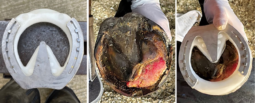
FIGURE 4, 5 & 6: A month after the first visit, Robbie Miller applied a 3rd Millennium aluminum heart-bar shoe attached to an Imprint shoe. Robbie Miller & Dr. Andrew Gray
Using what little wall there was at the medial heel area, I applied more thermoplastic to try to rebuild that area for medio-lateral balance. There was no way we’d get anything nailed on, so I used a 2-inch Derek Poupard HoofCast with therapeutic impression material over the heel bulbs to prevent abrasion over that area (Figures 7-9).
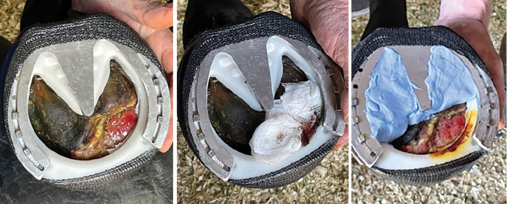
FIGURE 7, 8 & 9: Miller used a 2-inch Derek Poupard HoofCast to secure the package, while also adding therapeutic impression material. Robbie Miller & Dr. Andrew Gray
With supporting limb laminitis being a reality, the hospital staff was diligent in keeping ice packs on her off fore pastern. She also had a deep bed of pine shavings for full solar support.
After being at the hospital for about a month, she gave birth to a healthy, albeit small, filly. This little lady turns out to be quite a tough cookie, too. I know this from the solid kick I took from her on the thigh while taking a photo of mom’s foot.
At this point, Gray, Dr. Sonia Marcos and I feel that we can save this mare, and, with consent from the owners and stud farm management, we set about on an incredible journey with her. I daresay there were several times when we thought that we weren’t going to win here, but this mare and her foal kept us going.
Hoof Wall Resection
For the next several months, the veterinary team and I progressively removed the necrotic and granulating tissue and unattached hoof wall using hoof knives, half-round nippers and a cordless Dremel machine with an aggressive cylindrical bit.
On Oct. 7, we resected a window at the dorso-proximal (DP) area, which subsequently led to a full resection of the medial wall to the heel quarter at the depth of the connective area of stratum internum. The new, juvenile hoof wall was being compressed and therefore restricted at about 10 mm below the coronary band all along the dorso-medial area. There was also a lot of necrosis of the dermal lamellae, which created further infection concerns (Figure 10).
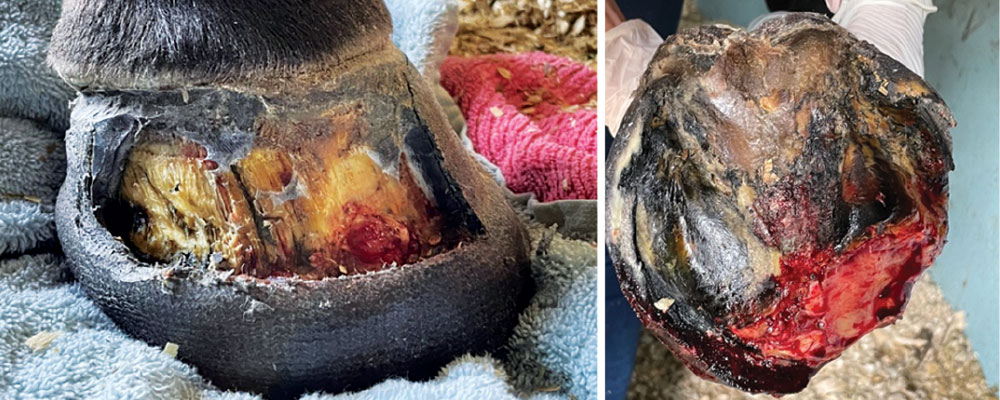
FIGURE 10 & 11: A dorso-proximal area of the broodmare’s hoof wall was resected. Subsequently, a full resection of the medial wall to the heel quarter was performed. Robbie Miller & Dr. Andrew Gray
A week later, on Oct. 14, there was a mass of prolapsed granulating tissue along the sole area which needed to be removed. We also removed a part of the dorso-lateral wall. At this point, the heels and frog were beginning to resemble a more normal appearance, which was encouraging. We fashioned a palmar sole support from impression material and thermoplastic that was attached with Elastoplast and Vetrap. The sole and dorsal area were medicated with an iodine solution (Figure 11).
I returned to the hospital Oct. 28 to check her out again. There was still an area just distal to the coronary band that was being restricted from growing down. I removed the last traces of old wall along the ridge of the new advancing hoof wall. Radiographically, there was still a lot of instability within the hoof capsule and P3 was clearly still rotated. From a DP view, there was still quite a significant medio-lateral imbalance. I reapplied the impression material and thermoplastic prosthetic and the hoof was re-medicated.
Remarkable Change
Mare and foal had been pretty much stable-bound the entire time. Gray was quite eager for them to get out to a paddock. On Nov. 11, after removing some granulating tissue along the dorso-distal border, medicating with sugardine and refitting her palmar support pad, they were introduced to some open space. Needless to say, the foal had an absolute ball being able to stretch her legs for the first time since birth. This was quite an emotional moment for all of us, as we had grown very attached to the two of them.
With most of the necessary hoof wall resection completed, it became a waiting game for the hoof to grow. She had been put on a course of Hoofmaker, made by the Irish company Thoroughbred Remedies Manufacturing (TRM).
In early March 2022, she presented acutely lame, and the alarm bells went off for obvious reasons. Marcos and I assessed her and found there was a subsolar abscess just medial to the apex of the frog. The area was opened for draining and a medicated glycerin poultice pad made by Farrier’s Friend was applied to the bottom of the hoof. Fortunately, she recovered within days.
Radiographically, the change was remarkable (Figure 12) and on March 24, I nailed a pair of shoes onto her front feet. On her near fore, I fabricated a wide bar using an old Legend Excel rasp and filled the palmar half with therapeutic impression material. Watching her walk out almost sound was an incredible feeling for all of us who had walked this long, uncertain road with this horse (Figures 13-14).
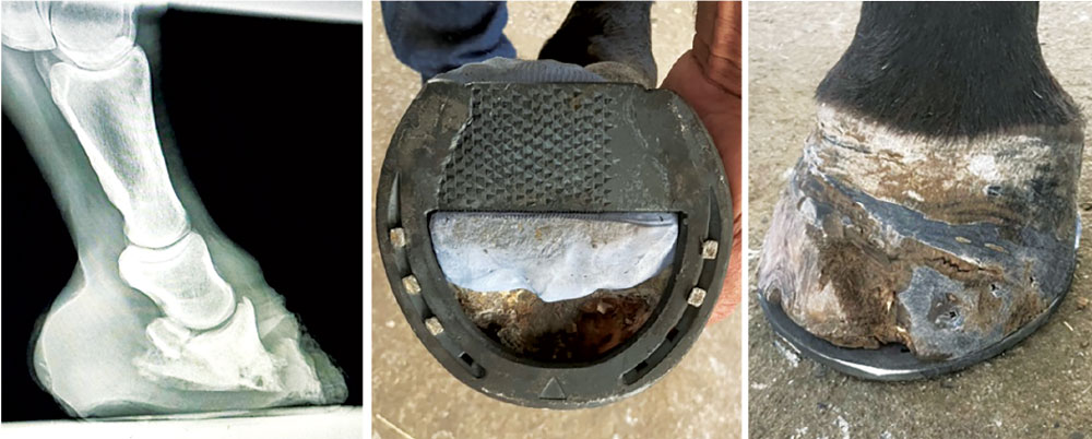
FIGURE 12, 13 & 14: A mass of prolapsed granulating tissue and a part of the dorso-lateral wall was removed. Robbie Miller & Dr. Andrew Gray
I took a drive out to the breeding farm to meet with the mare’s regular farrier and to shoe her once more on April 26. She was still slightly off on her near fore, but certainly still improving. Her hoof was growing out without issue and the capsular shape and integrity looked very good. She had good sole depth, and I was able to remove some excess hoof wall. The area where she had abscessed at the medial toe was callousing nicely, too (Figures 15).
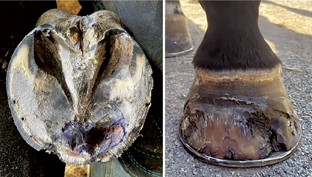
FIGURE 15: After a bout with a subsolar abscess medial to the apex of the frog, the mare recovered days later. Radiographic changes were remarkable. Robbie Miller & Dr. Andrew Gray
The fact that we managed to pull this mare through is a testament to the teamwork and commitment of an incredible group of professionals I am fortunate enough to work with. Add to that the faith of the owners and management of the farm that we are fully capable of saving their mare, so that she can hopefully breed them a champion (if she hasn’t already).