Farrier Takeaways

  • For issues like correcting a negative palmar angle, reliance on radiographs without the understanding the mechanics of the issue results in poorer footcare for the horse.
  • It is important for the veterinarian and farrier to discuss the cause and effect of cases, and to address the needs under their expertise.

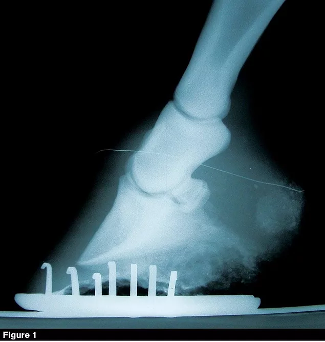
This year marks my 55th year of shoeing horses. Techniques have changed several times throughout that career based on subjective evaluation and misleading science. We now live in the age of information (and misinformation), and those who force their opinions on our farriery practices are expanding. Throw in a measure of “how-to” by owners and veterinarians who don’t shoe horses for a living, yet feel the need to tell us how to do it anyway. Fortunately, knowledge will help weed out the stupid stuff.

The autonomy that we should have to practice our craft is based on our experiences, skill and education. Actual decisions of normal hoof-care standards and the necessary techniques to address gait problems or lameness issues mediated by farriery should be left in the hands of the farrier.

Yes, input from other professionals involved with the case should be considered. However, the decision should be left with the farrier because that is the professional who will be the ultimate responsible party for those decisions.

LEARN MORE

Gain food for thought by reading other footcare observations by Randy Luikart at
AmericanFarriers.com/0720

Farriers have been known to make decisions that were not successful — some have made matters worse at times. We learn from both. Experience gives us the ability to identify when we are about to make a mistake and avoid doing it. When we are held responsible for those practices and techniques that have been imposed upon us by owners, trainers, veterinarians, massage therapists, etc., we tend to protest. That protesting can result in losing the client and straining relationships with all the other professionals involved.

Injuries, gait deviations or pathology evidence needing to be addressed by farriery techniques become a primary discussion point among us all. We vary our approaches based on past experiences, training, mechanical knowledge and the skill level required to accomplish the task. Let’s look at something commonly found, often not addressed correctly, yet still discussed regularly.

Influencing the Palmar Angle

Positive or negative palmar coffin bone angle is one of the most ubiquitous problems of the current times. Do we really understand all of the possible mechanics that occur both statically and dynamically that can allow this feature to become visible from a radiograph? Most of this discussion will center on the evidence found on the sagittal plane from lateral radiographic images (Figure 1 [top of page] and Figure 2).

Trigonometry — the study of right triangle ratios — was based on isosceles right triangles. Isosceles right triangles have one 90-degree angle and the other two are 45 degrees. That means the two sides of the right angle formed are the same length. The formulas derived from sides to angle ratios are applicable to all right triangles. These formulas then assist in providing a means to gather information in mechanics that is useful in calculating torque and associated tension to the soft tissue being subjected to that torque. Nature, being very symmetrical in design, has the coffin bone angle close to 45 degrees and if the pastern angle is the same, then you have the static tension on the two tendons defined. The tensions won’t be the same, as the superficial and deep digital flexor tendons (DDFT) have different conformation, muscle bellies, insertions and lever arms around the centers of rotation, but nonetheless, will be defined on that particular horse. You ask, “So?”

Go back to the positive angle finding on the sagittal plane indicated by the radiograph in Figure 1.What would you say if I asked you what possible mechanical applications would move the coffin bone into that position? There are two possibilities, not discounting the role of the shoulder in producing that effect. One is the tension on the DDFT/muscle unit and the other is vertical force component (weight of the horse) pushing on the extensor process in the direction of that vertical force from the pastern (steep) above the coffin joint. There are other dynamic forces of course, as the horse is not always standing still. Of primary importance for this discussion is the DDFT tension. There are hoof anatomy failures that will also yield this result but let’s remain on our thought process.

No matter why this muscle unit is being asked to fire and provide increased tension to the DDFT statically from a normal resting tension, it is indeed doing so. It is entirely possible that the mechanics of movement is also adding to it, but we will leave that complex discussion alone. The DDFT has two lever arms, one from the fetlock to the navicular bone; second, the arm created by the flexor surface of the navicular bone to the insertion on the base of P3. Each of these lever arms can provide tension. Those lever arms aren’t providing the increase at this point in time; the muscles in the forearm are. The tension each lever applies is based on the lengths and angles of foot and pastern during the weight-bearing phase of ambulation. So, if you assume that the static tension on the DDF muscle/tendon is the predominant force acting on P3, you would be correct.


It doesn’t make sense to address the problem by making the cause of the problem worse …


Radiographs are commonly pushed by the veterinary and farrier communities to resolve all aspects of farriery. Whether the center of rotation, balance around the coffin joint, or breakover, practically any aspect of our craft is being told, “If you make the radiograph appear more normal, it will fix the problem.” That assumption is absurd. Look at Figure 1. A higher positive angle is a result of increased tension on the DDFT. Farriers are being told to lower the hoof angle to put P3 in a more normal position so that a more normal-looking radiographic image could be obtained.

What does lowering the hoof angle do to the tension on the DDFT? Lowering the hoof angle always increases the DDFT tension. Lowering the angle of the hoof increases the distance from the flexor surface of the navicular bone to the insertion of the DDFT, increasing static tension from the distal end of the tendon/muscle unit — kind of like pulling on it from both ends. Now, lowering the hoof angle to make the radiograph look more normal might seem intuitively the right thing to do, but doing so will increase the tension on the DDFT/muscle unit. Lowering the hoof angle is a generally accepted procedure that’s routinely prescribed by veterinarians and also is supported by many farriers. Lowering the hoof angle has a linear relationship to the DDFT; it will always increase tension. If you proceed to lower the angle, then you increase both the static and dynamic influence on the strain of the DDFT.

It doesn’t make sense to address the problem by making the cause of the problem worse. Where does the common sense come into play? It really doesn’t. Those experts have fed us that line of fiction so long that perhaps they actually believe it. The mechanics that actually fix the problem may be just the opposite.

Take another look at the positive angle radiograph. Draw a line showing the angle of the base of P3, and without seeing the hoof, draw a line of the solar plane of the frog. It may be hard to do, but that is the plane you want to address. The anatomical connection of the frog to P3 is a good reference to realizing that plane. One must remember that the body of P3 has shifted within the capsule to produce our radiographic discrepancy. That shifting has produced stretching of toe lamina, rotational displacement of the lamina in the quarters and possibly shearing of the lamina in the heel area. That tells you the bone is not adequately connected to the hoof wall to utilize it to change that plane. Reread the previous sentence. Attempting to utilize the hoof wall to alter the position of P3 within is futile, as you have just increased the forces that have caused the positional changes in the first place.

Internal P3 positional landmarks vary during weight-bearing, that coupled with this increased instability, allows us the ability to modify those positional changes internally during weight-bearing. Methods may also be utilized for the dynamic assistance provided by farriery, but let’s just stick to trimming the hoof.

A lot of positive angled feet are dished, and a lot of negative angled hooves are bull-nosed. It is not necessary to dress them to a “normal appearance.” Addressing the mechanics within the hoof capsule will remove those deforming forces and the hoof recovers without unnecessary “shaping.” The bottoms of these two hooves are also distinctly different. The positive angled hoof has little cup to it and a short frog base, while the negative angled hoof has a longer frog and a much better cup. The goal is to restore the anatomical features that have failed in each hoof and allow it to deform the opposite way in a managed manner.

file name

The Approach is Lost

To address the lack of strength to the toe lamina in the positive angled hoof, simply leave enough wall length to cut an adequate cup. Establish a more normal cup than the one currently presented. Leave the frog tip alone (don’t go hunting for that infamous dot). If possible, you would like the frog to be supported by the ground or a frog plate/frog pad of some kind. I’ll explain this mechanical change. If the arc formed by the sole cup is to be efficiently supported, then building a vertical means to support the top of that arc is required. Cutting the cup and allowing the frog to bear weight at the tip does just that. The restoring of the cup and placing a vertical support within that arc will stabilize the hoof in the dorsal toe of the sole and reduce the tension on the toe lamina by minimizing the tip movement of P3.

You will want to leave enough frog for weight-bearing that the lever arm formed by the flexor surface of the navicular bone and the insertion of the DDFT is supported. Now simply cut the frog behind the area you have selected to bear weight to reflect the plane of the base of P3. The frog behind the area you have used at the tip should not bear weight and should float above the surface initially. You do not need to dress the outer wall.

Doing this will allow P3 to rotate slightly, heel first when bearing weight because the toe of P3 is not being allowed to drop further. This procedure reduces tension on toe lamina, reverses the rotational direction of the lamina in the quarters and will eventually help in the management of any shears existing in the heels.

Following and applying that same logic to a negative palmar angle P3 (Figure 2), the radiographs will tell you to raise the hoof angle. From that frozen speck of time in that radiograph, you certainly can build a case for doing just that — if your focus is to make the radiograph look more “correct.” Only after realizing that if the rotation of P3 is the other direction than a positive palmar angle, then the DDFT has less tension than is necessary to provide static stability. Raising the heels, applying wedge pads or elevating the hoof angle in any manner only reduces DDFT tendon strain further. Raising the hoof angle also has a linear relationship by decreasing the tension in the DDFT.

file name

Both of those accepted practices addressing negative and positive palmar angles will “make the radiograph look more appropriate/ideal.” But those of us who have followed those instructions only arrive at the next shoeing with the same problem, if not worse. It seems as though we have lost the common sense here as well.

The negative angle P3 is just the opposite of the positive angle hoof. The lamina is not stretched at the toe (actually being compressed slightly above the tip of P3), rotation in the quarters is just opposite of the positive angle P3 and the heels will be compressed rather than sheared in appearance. Management of that hoof problem is just the opposite of the positive angled P3. Cut the frog away at the front third so that it doesn’t bear any weight, remove enough foot so that little cup is formed rather than the deeper cup on the positive P3, and leave the frog at the heels to bear weight with the use of a frog plate or the ground. I have found that frog pads or frog plates are more effective, as not always do frogs bear weight on the ground with a shoe on. Figure 3 is the same foot in Figure 2 with a frog pad placed between the bars.

Doing that procedure will allow the heel of P3 to remain stationary when weight-bearing and the distal tip of P3 will now slightly rotate when weight-bearing. This will allow normal tension to restore to the toe lamina, change the direction of P3 rotation and the associate quarters’ lamina during weight-bearing and decompress the wall at the heels.

Each group of professionals has goals in mind of what success is. Trainers want to be able to race the horse Friday or show it this weekend. Veterinarians would like to quickly resolve the issue of lameness or address the artifacts that have been identified in their examinations. Farriers would like to see both — if it is possible. Discussions among these professionals will overlap, so resolving the issue might occur if the mechanical adjustments made actually remove the factors causing the problem rather than increasing them. Methods for mechanical resolution and prognostication of recovery are often the issues to discuss.

“Common sense,” I was once told, “is a fictional sense.” I have learned that common sense evolves from carefully analyzing a problem, taking that information and coupling it with the knowledge and skills necessary to provide a logical approach to solving that problem. In the case of positive and negative palmar angles, that approach has been lost.


July / August 2020