Pictured Above: Abscesses travel up to the coronary band where it’s easier to break through. If a horse doesn’t improve 1 to 2 days after blowing the abscess, the problem may be deeper.

Farrier Takeaways

  • When using hoof testers, a farrier has to be able to differentiate between pain vs. pressure. Start by testing the areas where the horse is not sensitive then examine the area that is suspect.
  • Don’t forget the limb opposite the afflicted foot. If the horse being treated is not bearing weight for 2 or more days, contralateral limb laminitis may occur.
  • If a horse suffers a puncture wound, do not remove the foreign body before consulting with a veterinarian. Take radiographs when possible.

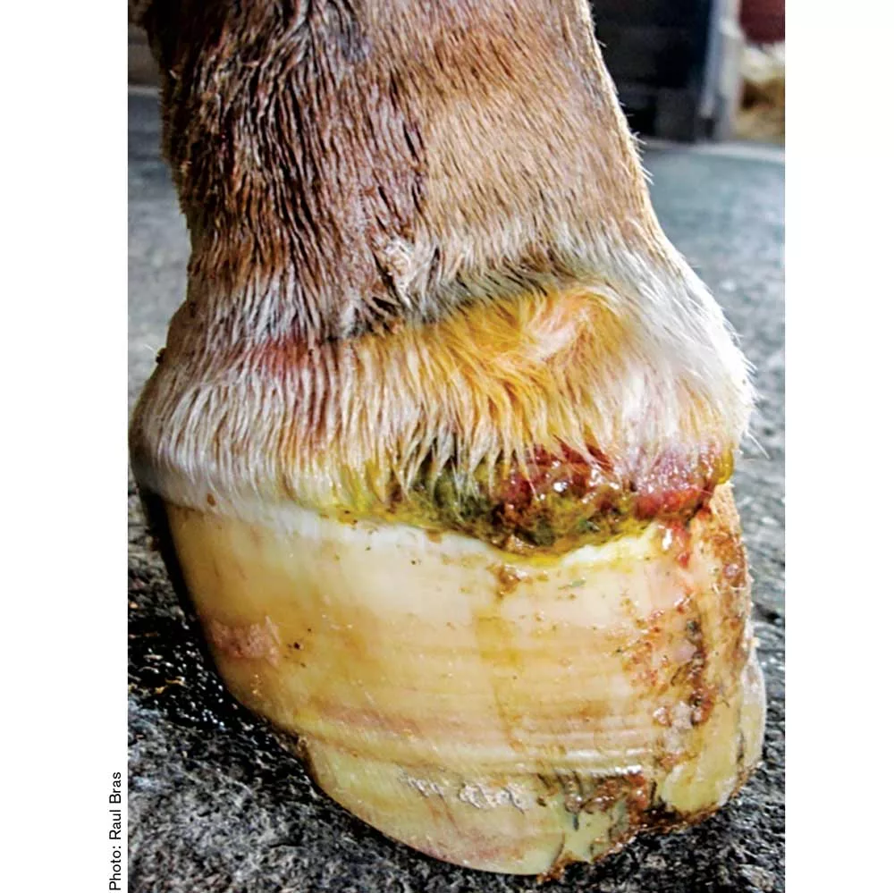
Foot infections are a common source of lameness in the horse. Understanding the causes of these infections and identifying their clinical signs are key to getting a horse back on the path toward health. Raul Bras, certified journeyman farrier and a veterinarian at Rood & Riddle in Lexington, Ky. discussed some of the different foot infections a hoof-care professional is likely to encounter, the importance of early identification and types of veterinary interventions that may be necessary at the Midwest Equine Podiatry Conference in Arlington, Wis. The conference was presented by Lodi Veterinary Care and Country View Equine Clinic.

Causes of Infections

Foot infections occur when a large number of microorganisms breach the protective barrier of the cornified hoof capsule — namely, the hoof wall, sole or frog — and invade the underlying sensitive tissue, Bras says. Infections can usually be attributed to one of four root causes:

1 Environment. Puncture wounds or gravel in which bacteria gain entry through defects in the white line.

2 Hoof capsule defects. Cracks, flares and contracted heels are breaches in the protective barrier.

3 Digital instability. Anything that causes displacement of the hoof capsule can predispose a horse to a pathology.

4 Systemic. Bacteria invade the foot through the blood stream through somewhere else in the body.

Talking to the client and understanding the history of the horse is where Bras gets some of his most valuable insight before examining the horse. Farriers who have had a horse on their books for a year or longer will be familiar with a horse’s history, but when it is a referral, asking certain questions can be instructive. Bras likes to know:

  • What day did it become acutely lame/how long has it been lame?
  • Is it unilateral or bilateral?
  • Does it have digital pulses?

Clinical signs of foot infection include lameness, swelling, heat draining tracks, increased digital pulses and evidence of hoof wall injuries. Assessing pain can be a challenge. When using hoof testers, a farrier has to be able to differentiate between pain vs. pressure.

“Is it just being a reactive horse, or does it truly have pain?” Bras asks. “If you feel like a horse is having lameness, you don’t just go to the apex of the frog and crack on it — because it is going to respond. Start in the areas where the horse is not sensitive then go to the area you are most suspicious about.”

After going through the process of learning the horse’s history, conducting a clinical evaluation, palpating the coronary band and checking digital pulse, if the farrier still can’t determine the cause, Bras recommends getting a radiograph. The views he typically requests are lateral, dorso-palmar and dorso-ventral. This can reveal an abscess or a fracture. Taking a systematic approach, and avoiding jumping to conclusions is in everyone’s best interest, Bras says.

“The last thing you want to do is give a lameness evaluation to horse that has a coffin bone fracture,” Bras says.

Abscesses and Puncture Wounds

The most common foot infection farriers are likely to encounter is abscesses. The “common” abscess scenario, Bras explains, is one in which the horse is sore, the hoof-care professional is able to locate the abscess, open it, drain it, pack the foot and in 2 or 3 days the horse is doing fine. Depending on the situation, a skilled farrier or a veterinarian may be able to successfully open an abscess, but the veterinarian will need to be involved to prescribe an anti-inflammatory and pain medication such as phenylbutazone, as well as antibiotics, as needed. The idea of withholding pain medication to monitor the horse’s pain progression is faulty logic, Bras says.

“It’s like if you go in for a root canal at the dentist and ask for pain meds and the doctor says, ‘No, I want to see how painful you are tomorrow.’ It’s the same thing with horses. If you open an abscess, get the horse something so it can heal a little bit quicker and not just be miserable.”

The key to abscesses is understanding how they travel through the tissue planes, Bras says. If a horse has sensitivity over the sole, opening up that spot on the sole would not address the underlying problem. It could, however, result in the corium swelling and seeping through the hole that has been made. Then every time the horse loads that foot, the hole will open and close, pinching the granulation tissue or corium, which will eventually lead to necrosis.

“There is a junction between the sole and the wall,” Bras says, “and usually it’s the white line. If there is sensitivity in the sole that might most likely migrate toward the toe I will go toward the white line and try to avoid any problems with the corium or granulation tissue.”


Is it just being a reactive horse or does it truly have pain ...


When the horse is going to blow an abscess from the coronary band, Bras may nerve block the horse and treat it with an overnight poultice comprised of a handful of bran mash to cover the hoof, povidone, dimethyl sulfoxide (DMSO) and Epsom salt combined with warm water to create a paste. He’ll then wrap the leg with a 5-liter IV bag and the overnight poultice and leave the horse for a day. When Bras checks in with his client the next day, oftentimes the client will report the horse is doing much better, which likely means the abscess broke through the coronary band. Bras then removes the poultice and has the client soak the horse and start treating it after it’s finished draining.

After actually opening the abscess and establishing drainage, it is important to keep the foot wrapped and protected from further debris for the next few days or the abscess will re-develop, Bras says. It may seem like common sense, but it’s important to communicate this information to clients. Bras also recommends monitoring the limb opposite the abscessed foot. If the horse being treated is not bearing weight for 2 or more days, contralateral limb laminitis may occur.

If the horse broke through the coronary band and for some reason doesn’t improve, Bras will re-take a radiograph to make sure the horse isn’t developing osteomyelitis or — worst case scenario, a sequestrum off the coffin bone. He may start doing regional perfusions rather than waiting for the infection to get into the bone. Regional perfusions provide a higher concentration of the antibiotic localized to the foot and are more effective than putting a horse on systemic antibiotics, but it’s not something all veterinarians are comfortable with, Bras says. Oftentimes, after 1 or 2 days on a regional perfusion, the horse will improve. At that point, Bras may deploy medical maggots to debride necrotic tissue and create healthy granulation.

If a horse develops a sequestrum, however, a surgical procedure will be necessary to remove it. However, this doesn’t mean general anesthesia and a surgeon are required. A skilled veterinarian may be able to remove the sequestrum with a local anesthesia while the horse is upright. Bras has been able to go in with a curate, non-invasively remove the sequestrum and clean the surrounding area. Veterinarians who attempt this have to be careful not to fracture the wing of the coffin bone in this process by being too aggressive.

Another environmental factor that is a common source of hoof infections is puncture wounds. Treat puncture wounds like they are your worst emergency, Bras advises. In many cases, the wound will be superficial and result in a subsolar or submural abscess. However, if the puncture wound is deep, an infection can occur in one of the many sensitive structures in the foot including the deep digital flexor tendon, the navicular bursa or the digital cushion.

LEARN MORE

Gain more insight about hoof infections by:

  • Reading “Identifying a Subsolar Abscess Isn’t Always Easy” in which Hall of Fame farrier Henry Heymering shares his tips for abscess detection.
  • Downloading the eGuide “Tackling Those Extra Tough Hoof Crack Concerns,” to take a more in-depth look a hoof capsule defects that can lead to infection.
  • Watching “Farrier Quick Takes” with Ohio Farrier Doug Hooper, to learn about one hoof-care professionals’ approach to using hoof testers.

Access this content by visiting AmericanFarriers.com/0320

A critical message to communicate to clients — whether horse owner, groom or barn manager — is not to remove the foreign body that has punctured the foot. Instead, Bras says, ask them to take a photograph of it and — if it’s not going to do more damage — leave it in the foot, but wrap it in a diaper or cotton bandage. The horse won’t stand on it because it is painful. Then, try to get a radiograph taken of the foot before the foreign body is removed.

“A nail might enter the foot at an angle,” Bras says. “You don’t know what direction it is going. A radiograph will give you an idea of what direction it is going and what structures could be inflicted.”

Other Causes of Infections

Although not an “infection” per se, hoof capsule defects are breaches in the protective barrier that allow bacteria access to sensitive tissue. Quarter cracks, false quarter cracks, hoof wall avulsions, chronic wounds, and quittor are a few of the hoof capsule defects that Bras has addressed in his career.

“Quittor confuses people,” Bras says. “It often is mistaken for a foot abscess because it breaks above the coronary band — on the skin. It’s basically necrosis of the collateral cartilage. In these cases, I might be a little more invasive because I have to debride the cartilage.”

The third most common cause of infections is digital instability. Farriers will most likely encounter this in cases of laminitis when the blood supply to the foot is reduced and the horse’s ability to fight infections is compromised, thus permitting abscess and necrosis to result.

If all other causes of infection are ruled out, the likely culprit is systemic, or when bacteria invade the blood stream.

The Farrier’s Role in Infections

The severity of an infection and prognosis of the patient will depend on the structures of the hoof involved. Infections of deep structures including the coffin bone, coffin joint, navicular bursa or collateral cartilages requires more care and veterinary intervention than infections of superficial structures. In all cases, the longer the infection goes untreated, the more damage the foot will suffer.

Identifying hoof infections early can greatly improve the outcome, Bras says. A farrier who sees horses on a regular trim schedule is in a key position to identify the lameness at its start, and partner with a veterinarian as needed to provide treatment. When the veterinarian and farrier work together, the horse is in the best position to make a recovery from a foot infection.


Learn More

Use Sugar to Treat Those Nasty Hoof Wound Injuries
The remarkable use of sugardine is now accepted more frequently by many veterinarians and farriers because of successful results and its economic significance.


March 2020