An infectious disease is one caused by an outside pathogen such as a bacteria, virus, parasite or fungus. Infectious diseases may or may not be contagious from horse to horse. Elsewhere in the body, infectious diseases are often the result of infection by one organism from a specific source. Examples include neurologic disease due to West Nile virus infection from the bite of a carrier mosquito or strangles caused by Streptococcus equi transmitted from a sick horse. However, the three major infectious diseases of the hoof capsule behave somewhat differently.

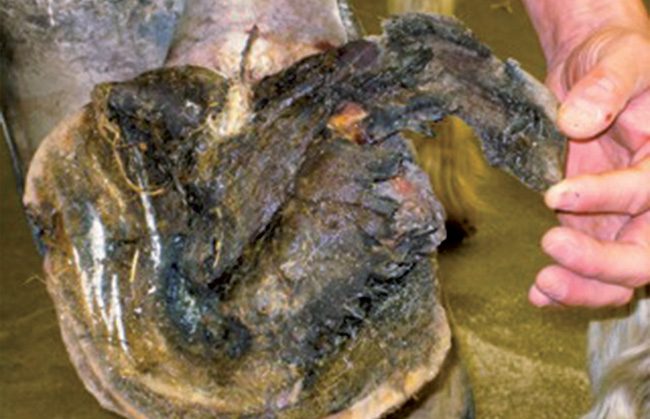
Thrush, canker and white line disease all appear to be caused by more than one organism. The organisms that have been isolated from each of these conditions are a mixed bunch and are generally widespread in the environment. So if the organisms are so common, why are some horses badly affected while others remain untouched? In most cases, infections of the hoof capsule appear to be “opportunistic,” meaning that some other condition — in the hoof or in the environment — has rendered the hoof vulnerable to invasion by organisms that would otherwise remain relatively harmless.

Thrush

The black, greasy, smelly frog sulci of a thrush-affected foot are familiar to most equine professionals. Thrush is defined as “a degenerative condition of the frog involving the central and lateral sulci, which is characterized by the presence of black necrotic exudates and a foul odor.” While thrush is generally thought to be associated with unhygienic conditions — high moisture, presence of manure and urine, mud or inadequate hoof cleaning — Stephen O’Grady DVM, MRCVS, of Northern Virginia Equine argues that thrush is unlikely to be found in a properly trimmed foot.

“If you keep the frog and the heels on the same plane, you don’t get thrush,” says O’Grady.

severe-thrush-after-debridement-showing-ddft.jpg white-line.jpg

Canker (above), thrush and white line disease (left and right) are examples of three different infections of the hoof capsule. Each of these behaves somewhat differently than the others. Farriers will often be the first to see the early signs of the infection.

In his paper “Background on Thrush and the Hoof’s Self-Cleaning Mechanism,” O’Grady describes the natural deformation and expansion of the sole and frog as the hoof contacts the ground. As weight is borne on the limb, “the third phalanx will descend, causing the sole to flatten. Descent of the coffin joint occurs as the navicular bone gives in a distopalmar direction, pushing against the navicular bursa and the deep flexor tendon, and finally causes expansion of the frog as it approaches the ground surface.”

According to O’Grady, in a foot with normal depth of the sulci, this expansion of the frog during weight bearing will remove accumulated material such as dirt and manure from the sulci.

Conformation Issues

No single organism has been identified as the cause of thrush; however, Fusobacterium necrophorum, an anaerobic bacterium that likes moist environments is commonly isolated.3 Deep and/or narrow sulci create an ideal opportunity for the colonization of the organisms that contribute to thrush.

According to O’Grady, hoof conformation “plays a big role” in the development of thrush. O’Grady identifies the upright, clubby, narrow foot with a long heel as the worst for developing thrush. In low-heeled horses, says O’Grady, those with “big frogs do well.” However, he states that “underrun heels with unhealthy frogs” are likely to come up with thrush.

Scott Morrison, veterinarian and farrier at Rood & Riddle Equine Hospital in Lexington, Ky., agrees. Morrison identifies “upright, boxy feet” as problematic and says, “Contracted heels tend to get thrush more easily.”

“As the central sulcus closes,” says Morrison, “you get a deep crevice, and you can get serious thrush, all the way into the digital cushion.”

While thrush is not typically considered to be a common cause of lameness, deep fissures — particularly at the heels — can cause pain, according to O’Grady. Once thrush is so severe that the horse becomes sensitive, it is critical to avoid compounds that may cause tissue damage, says Morrison. He warns against using strong, caustic chemicals such as formaldehyde, bleach and strong iodine in the treatment of thrush, saying that he has seen cases with severe chemical burns deep into the foot, even to the level of the deep digital flexor tendon.

Start With A Trim

For treatment, O’Grady first recommends a proper trim, taking the heels back to the plane of the frog if possible. Ideally, a rasp laid along the heels should touch the frog.

He also suggests removing the shoes for a month if the client will go along with it. He says this will allow the frog to come into contact with the ground, stimulating healthy frog growth. Damaged frog should be trimmed away, says O’Grady, and the frog and sulci cleaned with a wire brush. If a pain-causing fissure is present in the heel bulbs, O’Grady suggests using a straight bar shoe to restrict heel movement.

For topical treatment, O’Grady says most liquid antiseptics/astringents will work. He shies away from ointments, as he feels that they cause the foot to retain too much moisture. Instead he recommends painting the affected area with a mixture of Thrushbuster and rubbing alcohol, povidone iodine (Betadine) solution or a product called Real Heal.

For his part, Morrison recommends a mild disinfectant such as Betadine. He is concerned that many commercially available products may dry the frog enough to cause cracks, which may exacerbate the problem. Instead, he recommends using an oil-based antiseptic or painting the frog and sulci with alternating treatments of Betadine and pine tar.

Canker

While the early stages of thrush and canker may appear similar, the two diseases can be readily distinguished by their progression and a few key features. Both conditions arise in the area of the frog. But while thrush causes deterioration of the frog, canker is a proliferative dermatitis of the foot (proliferative pododermatitis.) Left to its own devices, canker will take over the foot like an aggressive cauliflower, invading not only the frog, but much of the rest of the foot as well.

“It starts at the heel bulbs and central sulcus,” says Morrison, “but then it can invade the sole, quarters and wall. I even had one bad case go up into the coronary band.”

canker severe-thrush.jpg chem-burn.jpg

Left to right: A hoof infected with canker being treated with medication. A hoof  that is badly infected with thrush. A hoof showing the effects of treating a hoof capsule infection with a caustic chemical compound.

Morrison and O’Grady agree that while canker may resemble thrush early on, the two conditions can be readily distinguished through close examination. The horse may not be lame during the initial stages of canker when only the superficial epidermis is involved.

The condition presents initially as a small area of granulation tissue (pink and granular looking) that bleeds easily when scraped. The focal area of granulation tissue will be surrounded by an area of light grey or brown tissue. It is in this early stage that canker may be confused with thrush; however, canker soon becomes proliferative, sending out lumps and fronds of abnormal tissue.

Due to this abnormal proliferation, canker often “outgrows its blood supply,” says Morrison. This lack of blood flow to the tissue causes necrosis, producing a foul smell.

Like thrush, canker cases appear to be more prevalent in moist conditions according to both Morrison and O’Grady. However, both practitioners agree that canker does not appear to be hygiene-specific, as both have seen cases of canker in well-maintained horses.

Cause Unknown

No specific cause has yet been determined for equine canker. While a species of spirochete bacteria has been isolated from canker lesions, the authors of that paper state that, “Given the different types of equine foot lesions in which spirochetes have been observed (canker, papillomatous pastern pododermatitis) the authors surmise that the spirochetes are opportunistic. However, this does not negate their possible contribution to the observed pathology either directly or in conjunction with other bacteria.”

While a similar condition, papillomatous digital dermatitis (hairy hoof warts) exists in dairy cattle and is believed to be caused by a spirochete species, no link has been identified between the condition of cattle and equine canker.

Although the clinical signs of canker are fairly distinctive, biopsy may be helpful in atypical cases or recurrent cases to rule out other conditions. Since samples tend to have a lot of surface contamination as a result of their location on the foot and mixed populations of bacteria tend to be found, culture is generally considered unrewarding.

Treatment of canker is best approached by collaboration between veterinarian and farrier. Given the potential degree of proliferation of canker lesions and the likelihood of hemorrhage as a result of excision, initial removal of the canker lesions should be considered a surgical task. Both O’Grady and Morrison recommend complete excision of the lesion, though they use slightly different surgical techniques.

In both cases, regional or general anesthesia and the use of a tourniquet are required. Morrison debulks the lesions surgically, then follows with a laser to debride and cauterize any remaining canker tissue. He then applies a shoe with a hospital plate to protect sensitive tissue. Morrison paints lesions with a mixture of metronidazole and tetracycline until the lesions begin to dry.

“I sometimes put bad cases on IV tetracycline as well,” says Morrison.

Morrison has recently found painting the site with a product called Tri-cide immediately before antibiotic application seems to help the lesions heal more quickly. Morrison stresses that he prefers to recheck canker cases weekly in order to monitor for regrowths that may need to be cauterized again.

O’Grady stresses the importance of thorough rather than aggressive debridement.

“If you cut everything out” says O’Grady, “you also take the good tissue.”

That slows the healing process. O’Grady also emphasizes the importance of “trimming the foot before anything else. The hoof wall at the heels needs to be on the same plane as the frog. If the frog is recessed, it will not heal.”

For hooves with damaged or atrophied frogs where it isn’t possible to take the heels back to the same plane, O’Grady will sometimes use impression material in order to engage the frog. “The frog needs to be in use,” he says. “It has got to be working as part of the heel.”

O’Grady follows surgical excision with cryotherapy (freezing). He feels this approach knocks out any residual organisms in the area. O’Grady dresses the debrided lesions with a mixture of 10% benzoyl peroxide in acetone with crushed metronidazole tablets. Recently, O’Grady has seen some benefit to systemic therapy with prednisolone as well.

While O’Grady reiterates the importance of proper trimming to the success of canker therapy, he states that, “Canker should be treated in conjunction with a veterinarian. This is not something a farrier should be doing alone.”

White Line Disease

Like canker, white line disease appears to be a condition of opportunity. In both conditions, O’Grady feels that affected horses must have “a propensity to come down with it,” since one horse in a barn may be affected while the others remain fine.

Also called seedy toe, yeast infection, hoof-wall disease, environmentally induced separations, onychomycosis and Candida, white line disease results in progressive separation between the stratum medium and stratum internum of the hoof wall. Unlike laminitis, the sensitive tissues are not typically involved.

In one study, scanning electron microscopy (SEM) analysis of hoof samples with horses showing clinical signs of white line disease revealed “severe alterations of the horn structure in horn samples infected with keratinopathogenic fungi compared to horn samples from a sound hoof. The most evident changes were deterioration of the tubular structure of the horn wall, disruption of the horny layers, superficial lysis of cornified cells and the presence of fungal elements.”

In this study, the authors identified fungal elements associated with damaged hoof in each case. However, as they acknowledge “this does not prove causality, as already damaged hoof horn might be more prone to fungal infection than sound hoof horn.”

Generally, the infectious elements (bacteria and fungi) that have been isolated from cases of white line disease are considered to be opportunistic pathogens. White line disease always occurs at areas of separation of the hoof wall.

“We don’t know what causes the separations,” says O’Grady, “But you can fill hoof wall separations with anything in 10 horses (and nothing will happen), but the 11th will progress to the coronet. The horse has to have a propensity (for white line disease.)”

In fact, O’Grady says that it has not yet been possible to experimentally transfer white line disease from one horse to another.

Morrison agrees with the model of white line disease as an opportunistic condition.

“We see white line even in the best kept horses,” says Morrison. “It’s more a mechanical problem than anything else. The fungus (that has been isolated from affected hooves) is everywhere. They’ve even found it on fruit in the supermarket.”

Like O’Grady, Morrison feels that the common nature of the organisms found in white line disease indicates that affected horses must have some predisposition that renders them susceptible to infection. However, to be on the safe side, he does say that if a farrier is working on a horse with a significant case of white line disease, he would recommend “cleaning and sterilizing your tools before going to another foot, and disposing of the shavings — not just sweeping them into a stall.”

Both Morrison and O’Grady agree that barn hygiene seems to have little bearing on the risk of white line disease. In fact, Morrison feels that he sees more cases in stalled horses (even in well-cleaned barns) than in horses on pasture. Morrison also sees more cases in shod horses.

“I’ve seen a few cases in barefoot horses, but I do think it is more common in shod,” he says.

While O’Grady believes he has seen more cases of white line disease in shod horses, he admits, “it is hard to say.” He does note that with an early separation, “you can take a loop knife, create a trench to good tissue, put a shoe back on and 4 weeks later, it will have advanced. However, you can take that same trench, fill it with putty, and it heals.”

Severity Varies

White line disease can occur at areas of separation anywhere along the white line. Early signs of white line disease include white, chalky, powdery areas at the sole-wall junction and widening of the sole-wall junction. As the diseaseprogresses and the hoof separates more proximally (toward the coronary band), tapping on the hoof wall with a hammer may produce a hollow sound.

“There is no predictive factor for severity,” says O’Grady. “But, it can progress if you don’t address it. Don’t ignore the early signs.”

As the disease progresses, O’Grady notes that the hoof wall begins to lose attachment, particularly at the toe, and the the sole drops.

“If you look closely around the outside wall, you see a little bit of dish at the toe. If the separation is too extensive, you get displacement,” he says.

Morrison stresses the importance of monitoring the shape and structure of the foot. “You want to minimize hoof capsule distortion,” he maintains. “Hoof capsule distortion equals an entry site.” He notes that separations are more likely to be found at the toe in “clubby” or laminitic feet, but will appeaer in the quarters near the heel in a low-heeled horse.

White line disease also invades damaged areas of the wall such as abscess tracts, according to Morrison. He feels “some feet seem more susceptible.” Among these, he lists hooves with “dull walls, brittle feet.”

For these at-risk feet, Morrison often recommends hoof supplements or dressings to protect the wall. He likes “pine tar on dull, brittle feet. It seems to keep the moisture content stable. It has natural anti-fungal and anti-bacterial properties.” However, he cautions against some commercial products, which he believes can “make the feet too soft.”

Both Morrsion and O’Grady emphasize the importance of proper farriery in managing white line disease. Early on, in cases of hoof capsule distortion, such as long-toed, flared or dished hooves, Morrison suggests that farriers can “take tension off of the dish by rockering the toe. You want to keep the white line from getting more stretched.”

If the area of separation is extensive enough to warrant hoof wall resection, O’Grady cautions that it is important to “do the farriery first” and to make a plan.

“Figure out how you can attach something before the resection. Once you have a plan, then do the resection,” he says.

The goal of shoeing following a hoof-wall resection is to unload the affected area, says O’Grady.

In debriding areas of hoof wall damaged by white line disease, it is important not to leave any pockets of undermined wall. Even though substantial amounts of hoof wall may be resected in severe cases, O’Grady feels that resorting to glue-on shoes is unnecessary.

“You can use clips and screws,” he says. “Be creative. All it is, is having a bit of skill.”

O’Grady feels that he may be seeing problems with some of the glue-on and heat-producing polymer compounds in white line-affected hooves. Additionally, though resected hoof wall may be unsightly, “patching” the damaged wall with acrylics or glue is not advised, says Morrison.

While Morrison likes to use Clean Trax for the post-resection treatment of white line disuse, O’Grady feels that once the damaged tissue is properly debrided, no chemical treatment is necessary. He will occasionally use a disinfectant such as Betadine as a dye marker to follow tracts for further debridement.

Ultimately, the farrier is the first line of defense. Early warning signs of hoof capsule infections such as thrush, canker or white line disease are likely to become first apparent during hoof care. Prompt recognition of these early signs and action by the farrier either through appropriate trimming or alerting the owner to the need for veterinary attention can make a difference in the outcome of these cases.


Learn More

Use Sugar to Treat Those Nasty Hoof Wound Injuries
The remarkable use of sugardine is now accepted more frequently by many veterinarians and farriers because of successful results and its economic significance.