
American Farriers Journal
American Farriers Journal is the “hands-on” magazine for professional farriers, equine veterinarians and horse care product and service buyers.
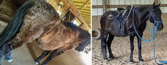
Relevante displays hypertrichosis, or long hair coat, which is often a clinical sign of pituitary pars intermedia dysfunction (PPID). Following treatment, Relevante coat returns to normal. Photo by: ECIR
It wasn’t that long ago that a diagnosis of Cushing’s disease — Pituitary Pars Intermedia Dysfunction (PPID) — was felt to mean the horse had less than 5 years to live. The cause of the laminitis in these horses was poorly understood and, therefore, poorly managed. We know better now.
The Equine Cushing’s and Insulin Resistance Group was founded in 1999 by an owner with an atypical Cushing’s mare who wanted to promote the dissemination of correct scientific information. We have followed the research closely, as well as thousands of cases. I can’t think of any situation where it’s more critical for owner, veterinarian and hoof-care professional to be fully informed and working together than in laminitis care.
We often describe the newly diagnosed laminitic horse as “acute.” What does acute laminitis mean?
With the possible exception of laminitis of pregnancy, few endocrinopathic laminitis (EL) cases are truly acute, where acute means the day before they…
American Farriers Journal is the “hands-on” magazine for professional farriers, equine veterinarians and horse care product and service buyers.

American Farriers Journal is the “hands-on” magazine for professional farriers, equine veterinarians and horse care product and service buyers.
Download these helpful knowledge building tools

We are here to support you.
We stock a wide range of high-quality products from trusted brands to ensure durability, performance, and reliability in every job you undertake. Our extensive inventory of horseshoe products and farrier tools means you can find everything you need in one place, saving you time and effort. Your satisfaction is our top priority. We are committed to providing excellent customer service, prompt shipping, and hassle-free returns.