The email arrived in Mike Bagley’s inbox on May 6, 2020. The horse owner, who didn’t know Bagley, inquired about whether the Canton, Ohio, farrier would take on her aged Standardbred gelding with white line disease.

She provided photos of Andrae’s right front hoof, as well as contact information for her veterinarian. At a glance, it looked like the horse only had vertical cracks (Figure 1). The horse owner’s regular farrier didn’t realize the cracks indicated white line disease. However, radiographs revealed a large void in the hoof wall (Figure 2). White line disease had invaded at least halfway up the wall and the hoof had a thin sole. It turned out to be the second worst case of white line disease that Bagley has seen.
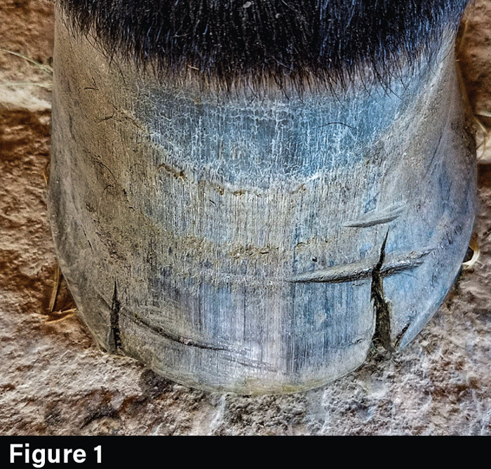
Hoof-Cracks

Andrae’s right front hoof presents with vertical cracks.
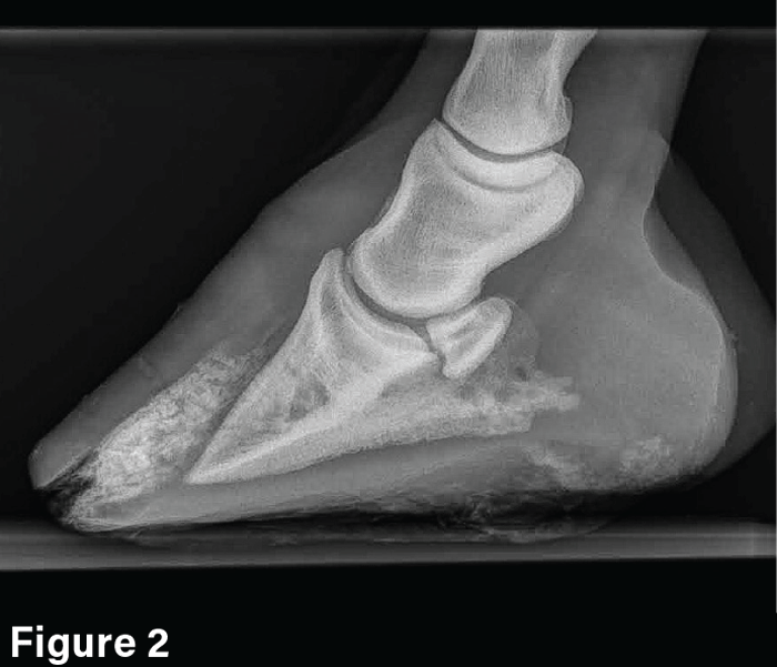
XRAY

A radiograph indicates that white line disease invaded at least halfway up the hoof wall, as well as little sole depth.

“I called the vet and he said he believed I needed to resect a good amount of the hoof wall but that he wasn’t sure what it would entail,” says the vice president of the International Association of Professional Farriers. “I explained it to him. He agreed that sounded good and turned me loose.”

Bagley asked the vet if he would like to be there to perform a nerve block or to respond in case there was too much bleeding.


“The outcome was successful because of the teamwork between me, the veterinarian and the horse owner…”


“He said I wasn’t going to get into anything sensitive,” Bagley recalls, “just that I needed to take off a bunch of hoof wall, and he was right.”

First Visit

Bagley, CJF II, APF-I, scheduled his first appointment for June 6 and invited fellow Ohio farrier Joe Rindchen, APF-I, to ride along and assist. Rindchen was also interested in therapeutic work. Bagley saw it as an opportunity to share his resection knowledge and teach a young farrier how to perform one.

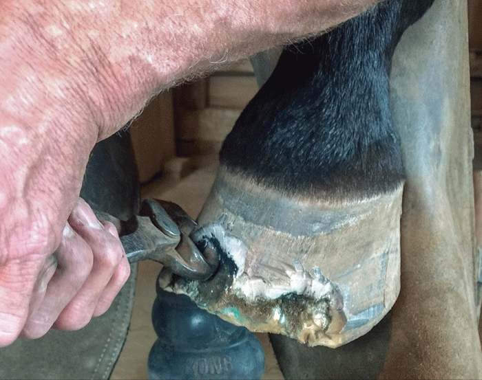
The gelding’s toe was about 3 ½ inches long when the pair arrived. Bagley resected the wall up about 2 inches. He chose to use half-round nippers because they cut into the loose hoof wall more than flat nippers would allow (Figure 3). Then he used a Dremel tool to smooth the rough edges (Figure 4).

Hoof-Wall

Mike Bagley resects the hoof wall with half-round nippers. Half rounds are more efficient in completing the task than flat nippers.

Dremel-Detail

Bagley uses a Dremel for detailed work during the resection.

Bagley carefully balanced resecting enough of the hoof wall to promote healing while taking precautions against removing too much of the hoof (Figure 5). When too much tissue is removed, a horse can become lame, and its hoof structure can be compromised. The structure around the foot still provides protection and stability even though the walls of the hoof are no longer connected to where the defect occurred. In extreme cases when the dorsal wall is drastically resected, all connection from the medial to lateral is lost. That can compromise the horse’s long-term soundness and increase the likelihood that the remaining hoof capsule will fail.

Resection

Bagley removed about 2 inches of hoof wall during the resection. His goal was to remove enough wall to promote healing while taking precautions to avoid compromising the hoof structure.

After the resection, he applied an Ironwork heart-bar shoe for support and shod the left hoof with a keg shoe to help keep the horse balanced. Bagley chose the heart-bar shoe because the horse had a thin sole and the hoof wall was extensively compromised.

Farrier Takeaways

  • Knowing the client’s goals for treatment and having a horse owner who is willing to manage the horse based on recommendations helps in deciding which course of action to take.
  • Working in partnership with a veterinarian leverages the expertise of both practitioners and gives a complete picture of how advanced a case of white line disease is with radiographs.
  • Inviting farriers along on cases offers a hand to help for the day, as well as an opportunity to share your expertise with other farriers.

The vet had already advised the horse owner that management on her part was critical to healing the hoof. He instructed her to keep the horse in a clean, dry environment. Given the frequently wet conditions in Ohio, that meant keeping the horse in a stall and using powdered lime to disinfect the space. The gelding only received turnout on dry days, and fortunately for Bagley, she was on board with providing the care the horse needed to fully recover.

“I left the client with copper sulfate crystals and instructed her to mix 1 tablespoon into a cup of white vinegar to make a spray to apply daily,” he says. “The white vinegar is a driver and drying agent, and the copper sulfate is anti-bacterial.”

Follow-Up Visits

Bagley returned for a follow-up on July 25, and while the hoof was looking better, he could still feel voids between the layers of the hoof walls (Figure 6). He was not sure how much more of the attachment was compromised, so he resected about another ½-inch and reset the heart-bar shoe. On subsequent visits, Bagley could see the hoof was improving, but the gelding continued to throw the heart-bar shoe.

Voided-Hoof-Wall

During the July 25 follow-up appointment, the hoof looked improved. However, Bagley felt voids between the layers of the hoof wall. He removed another half-inch of the hoof wall and reset the heart-bar shoe.

During the September visit, Bagley could see that the horse had grown some sole and that the attachment was solid (Figure 7). However, he still wanted the extra support of a bar shoe and made the straight bar to replace the heart bar that kept coming off.

Sole-Growth

The horse presented improved sole growth and solid attachment during the Sept. 5 follow-up.

“I used a 5 slim blade nail to poke up between the layers of the hoof wall (Figure 8),” he says. “Then I used the Dremel to remove just a little more.”

Probing-Hoof-Layers

Bagley uses a 5 slim blade nail to probe between the hoof wall layers. Bagley used a Dremel to remove more infected hoof wall.

By October, the hoof was growing out to the point that the hoof wall had a solid attachment throughout the structure. Bagley performed one last debridement to the remaining affected area (Figures 9a and 9b). On subsequent visits between October and May 2021, Bagley confirmed the void disappeared (Figure 10) and the hoof continued to grow well — so well that by May the resected area was nearly grown out (Figure 11).

Debridement

The hoof continues to grow nicely with solid attachment during the Oct. 17 follow-up. Debridement is necessary, but it will be the last.

Bar-Shoe-Reset

No voids were found with a nail during the Nov. 28 visit. The hoof continues to grow nicely. The bar shoe was reset.

Grown-out-hoof

The hoof is almost grown out during the May 6 follow-up appointment.

“It took a little over 9 months to completely clear up,” he says. “On my last visit — almost exactly a year later, his hoof was completely grown out (Figure 12). I applied keg shoes to protect the hoof and make sure everything stayed together.”

Keg-Shoes

During Bagley’s last visit on June 18, the hoof is grown out. He applies keg shoes.

Return to the Original Farrier

On Aug. 8, the horse’s regular farrier resumed trimming and pulled the shoes as the horse was sound barefoot (Figure 13). Ultimately, the horse owner’s goal was to get the horse pasture sound, which was accomplished.

Barefoot--sound

Andrae’s regular farrier resumes trimming and shoeing on Aug. 8. Andrae returns to being barefoot and sound.

Since the horse was not a regular client, Bagley is unsure what initially triggered the white line disease in this horse. It didn’t appear to be the environment — two other horses lived on the property and did not have it, which is not unusual. In addition, the horse only had it in one hoof. He believes the cracks in the hoof may have been the root cause.

“Sometimes what happens is that a hoof wall gets overgrown and cracks,” he says. “Bacteria start invading, but I’m not really sure on this one.”

White line disease does not always require veterinary intervention. It is, however, important to work closely with the horse’s veterinarian when the infection is aggressive. It’s also necessary for the diagnosis of underlying pathologies. Radiographs can reveal the extent of the disease. A comprehensive analysis of the hoof structure can also reveal other pathologies that could contribute to the disease.

“Having the vet open to a partnership really helped the horse,” Bagley says. “I work with some vets in my area and some I refuse to work with because they want to hand a prescription or say try this or that without asking me if there is a better way. This guy was really good, and it made all the difference for the horse. The outcome was successful because of the teamwork between me, the veterinarian and the horse owner all doing their part.”