
Shooting X-rays isn’t as simple as pointing the beam at the hoof and snapping an image. After all, it’s critically important to veterinarians, farriers, horse owners — and the horse — that radiographs contain accurate information.
Brent Barrett, an Ocala, Fla., veterinarian and an American Farrier’s Association Certified Journeyman Farrier, follows a list of tasks that he accomplishes to capture a clean radiograph.
1. Clean the feet. Debris on the hoof wall and in the bottom of the foot can provide misleading information.
2. Even feet placement on blocks. “Does it make a big difference if one foot is on a block and the other isn’t?” he asks. “I haven’t studied it much, but it’s just a good habit. You don’t want to guess if the subtle changes are an issue. You don’t want to blame it on them being shifted because of some imbalance being on blocks.”
3. Horse stands square and head/neck straight. A horse that stands square and straight ensures that its posture is true. Otherwise, incorrect information, such as uneven joint spacing, could be captured.
4. Mark coronary band and dorsal hoof wall. “I mark the coronary band with barium paste,” Barrett says. “I start my mark where you can just feel over the ledge of the coronary band and the hoof wall. If you’re off by 2 or 3 millimeters on a laminitic horse, that may be crucial because you need to know if the horse is sinking.”
5. Mark tip of frog. “If you want an internal to external reference point to determine where to put breakover or where the tip of the coffin bone is, the tip of the frog is a good place to go,” he suggests. “The veterinarian can measure that for you if you’re not together or you don’t have access to the radiographs.”
6. Beam location. When radiographs to assess balance are necessary, Barrett encourages the farrier to speak up.
“You have the right to say,” he says, “‘Can we do this a little differently?’”
To achieve this, the center of the X-ray beam should be focused on the ground surface of the hoof.
“The X-ray beam is like a shotgun,” he explains. “It comes out and spreads. Whatever you’re looking at, whether it’s a joint or the foot balance, you want the beam passing straight through it.”
7. Plate placement. Barrett wants the plate as close to the foot as possible to reduce magnification.
8. Block markers. Barrett wants to see only one coffin bone wing, one branch of the shoe (Figure 1a) or the correct block marker. Since he made his blocks, Barrett placed four screws within them.
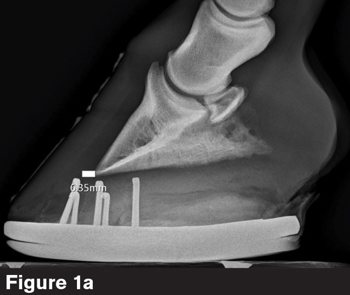
“The screws are set at the same plane,” he says. “When I see two screws instead of four, I know I’ve shot at the top surface of the block, the foot surface, or the ground surface of the shoe.”
When an image captures two coffin bone wings (Figure 1b), one of three things could be occurring.
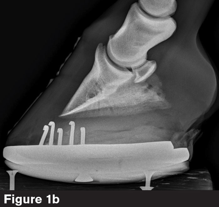
“You can have an asymmetrical coffin bone within a foot that’s actually balanced,” Barrett says. “You might have a medial-lateral imbalance. Or your beam is passing at too much of an angle underneath the bottom of the coffin bone.”
When two coffin bone wings and four screws are visible, as seen in Figure 1b, anatomical accuracy is in doubt.
“How do we measure sole depth?” he asks. “If the sole is superimposed by a wing of the shoe, we don't know where it ends. If you’re measuring the PA, a lot of times it will be different from one wing vs. the other. So, it’s crucial to get the soft tissue parameters on the bottom of the foot flat.”


