Farrier Takeaways
- Identifying the earliest onset of any change in exterior hoof health can be advantageous in the outcome.
- Once a decline in overall foot health has been noted, hoof-care providers can be led down a path of reactive footcare in the hope to regain the structural function of the capsule.
- The application of therapeutic support can bring significant value and comfort to the horse. However, without considering the primary onset, the condition may continue to arise if the primary cause isn’t identified.
As equine hoof care providers, we are often tasked with the challenge of interpreting the hoof’s response to neglectful husbandry or the horse’s adaption to load-bearing or pain.
There can be a multitude of factors that ultimately change the morphology of the horse’s hoof capsule. These variables can be difficult to assess and navigate as the nuances around every case can be completely different. Footcare may need to be addressed in a variety of ways to resolve different case presentations.1
Identifying Changes
In many ways, the horse’s foot is an amazing piece of bioengineering. The complexities of the internal mechanisms of perfusion, energy distribution and locomotion are all intricate pieces of the puzzle of overall foot health.2 Many of these internal structures also provide detailed services to the overall external health of the foot.3 For hoof-care providers, identifying the earliest onset of any change in exterior hoof health can be advantageous. It allows the hoof-care provider to start to make necessary changes and communicate what we are seeing to the appropriate people.
It can be easy to become focused on certain parts of anatomical structure when you see a decline or advancement in foot health. And, although at times there are isolated areas of interest, we have to consider that the hoof capsule works with some integration or unity of each and every structure. Therefore, when we are assessing each foot, the overall health of the hoof should definitely be considered.3
The hoof capsule’s ability to manage locomotive strain could be considered a foundation for all footcare. The hoof’s capacity to adapt to external force can be appreciated in shape, function and mass. Once a decline in overall foot health has been noted,1 this can lead hoof-care providers down a path of reactive foot care in the hope to regain the structural function of the capsule.
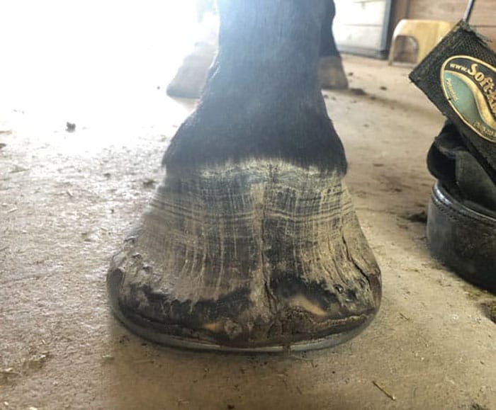
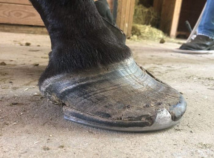
Full-thickness toe cracks accompanied the hoof distortion. This toe crack was unstable, meaning the hoof capsule was in two halves. Stabilization of the crack was needed.
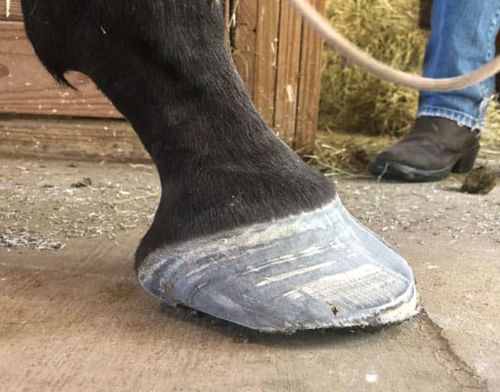
First trim: The dorsal wall was brought back significantly by removing the wall at the origin of the distortion. Note the prolapsed sole indicated by the shadowing under the hoof.
The management of peak force during locomotion could be considered a priority in today’s equine athletes. Initial contact, mid-stance and breakover can be segregated4 and, as hoof-care providers, can allow us to get very specific with our shoeing intention for each and every phase of the loading sequence. It could be entirely possible that each phase of ground contact might need careful consideration and management for horses that compete at a high level.
The horse’s locomotive tendencies and the foot’s ability to withstand the loading pressures will often be visible in the function and form of each limb and foot. The deformable nature of the hoof capsule will, over time, give the hoof-care provider feedback on the overall performance of the structure of the hoof.1 Promoting better foot quality can often lead to the hoof capsule performing better and allowing the structures to stay within the confines of their physiological limitations.
LEARN MORE
Gain more insight from Stuart Muir at
AmericanFarriers.com/0322
Hoof mass could be considered just as important as hoof balance. The epidermal structures of the hoof capsule offer considerable protection against trauma-based injuries. Blunt force trauma or excessive concussion caused through locomotion potentially has the ability to alter the hoof’s growth mechanisms.5 The hoof capsule’s response to extended periods of locomotive strain often results in reduced hoof capsule growth. When the hoof capsule is perfusing poorly, we could only assume that the structures that lay internally could also be damaged or functioning outside of their physiological scope.2
One of the largest concerns when attempting to rehabilitate feet is the internal and external composition of the structures we are trying to target. While there is not a lot of literature on the relevance or connection between the internal and external mechanisms, most hoof-care providers have noted that some lameness and hoof capsule distortions seem to rehabilitate faster and have more longevity than others. Therefore, as hoof-care providers, we may have to consider the viability of the internal and external structures we are trying to positively influence.6
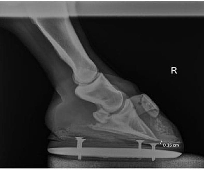
Radiograph after the first shoeing. Showing minimal sole depth.
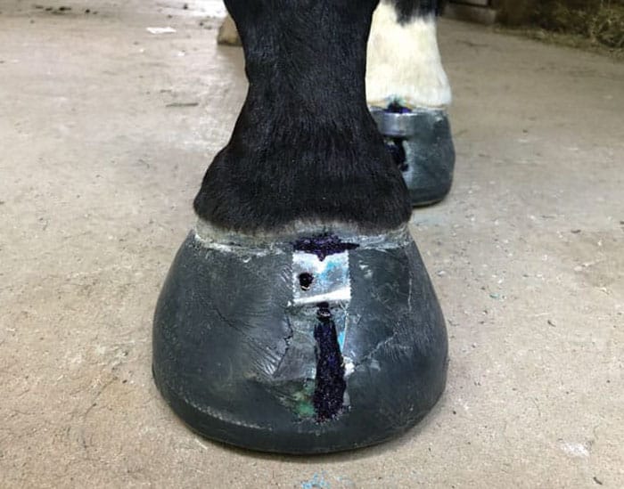
Careful attention to trimming and shoe selection was important to the success of this case. Due to the lack of wall structure and the lack of sole depth, an indirect shoe was glued on. The sole region had a very soft impression material inserted between the foot and the base of the shoe. The density of the impression material was important. I wanted to add support but I didn’t want to create pressure across the sole region.
Encouraging Robust Feet
One of the most direct approaches to having good-footed horses in your clientele is to be pragmatic and ensure that the horses you are working on have been encouraged to develop good, robust feet. A foot with a good external structure can be invaluable to the horse and aid in the dissipation of the loading forces acquired during locomotion.3 The external characteristics of the hoof would ideally be distortion-free and possess enough density to allow the structures to operate within the physiological range they were intended for. A well-developed foot will service the horse well into retirement if the horse is managed correctly.
The wall structure of the horse can be evaluated in terms of strength and health. Thin walls have been thought to reduce the effectiveness of the function of the structure.1 Divergent growth rings can also indicate that the horse has undergone some element of metabolic stress2 and the location and depth of the rings can offer a timeline of the event. The wall structure is also composed to assist in the dissipation of force during locomotion.5 The outer layers of horn are dense and hard while the inner layers of the stratum internum are softer and allow for the concussion to be reduced as the vibrations migrate toward the bony structure. Healthy wall composition will be uncompromised, thick and appear slightly glossy on the most outer layer.
The delivery and return of blood to the capsule could be considered another aspect of the fundamental features that assist in the reduction of concussion during locomotion. Perfusion of the equine limb is delivered and returned via a complex arrangement of arteries, capillaries and veins that descend and ascend the horse’s limb and foot via the neuro-vascular bundle.7 The delivery of blood supply to and from the hoof is probably hydraulic in nature and made possible by centrical force caused through the swing phase of locomotion and dissipating force caused through ground contact. As the blood enters the foot it supplies the foot with nutrients, oxygen and glucose.
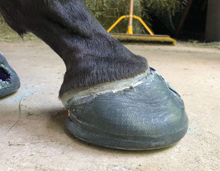
Lateral view after first shoeing.
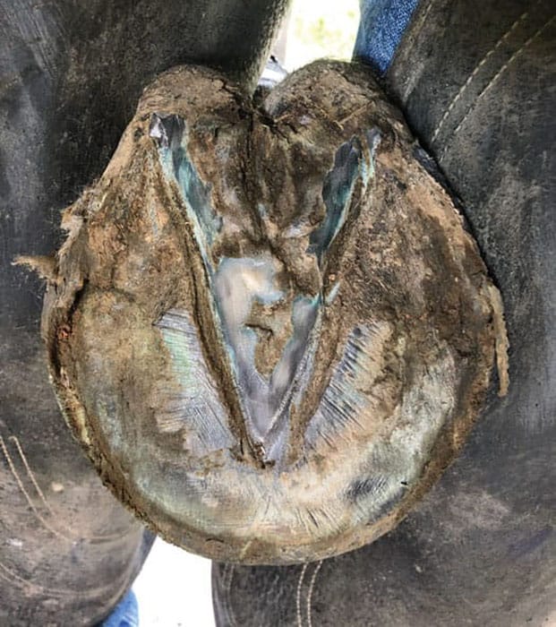
Second shoeing: The foot is slowly starting to grow some sole mass. This is an important factor in this case. Without the return of good quality keratinized sole mass, the sole structure wouldn’t have the ability to provide its fundamental role of offering the coffin bone protection. Without sole mass, the foot would be vulnerable to bone bruising and inflammation.
Ideally, the hoof’s internal structures would be uncompromised and allow full perfusion in all areas of the capsule.
It’s the author’s opinion that it is possible that we could experience regional deficits of blood within the hoof capsule that are influenced by proximal and internal loading pressure changes.8 When hoof mass is missing within one or more quadrants of the capsule, it can be useful to reevaluate the limb conformation and limb balance to decipher the origin of the cause. Regional or widespread loss of sole mass could be considered an indication that the dissipation of force or ground reaction force is stressing the hoof capsule beyond its physiological limitations in that area, and the underlying structures are excreting less new horn.
The condition of thin soles has been noted in numerous farriery publications as a precursor to lameness. The condition is often diagnosed by the sole region being easily deformable under manual palpation and accompanying positive pain response. Underlying regional pain is often isolated around the apex of the coffin bone or, alternatively, the palmar or plantar processes.1
The sole contour of the hoof capsule would ideally mimic the solar surface of the coffin bone with the addition of sole mass. A foot with a healthy, well-suspended arch offers protection from trauma-based injuries from uneven ground contact. The solar arch can be affected negatively by a reduction in sole mass, trauma, environmental changes and body composition changes. In cases in which the solar arch has become flattened or fails to provide sufficient protection to the internal structures, bruising or inflammatory conditions may become possible.3
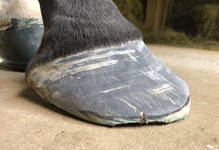
Second shoeing: The impression material has done a nice job of offering sole support. We are seeing a significant improvement in the sole position and foot quality.
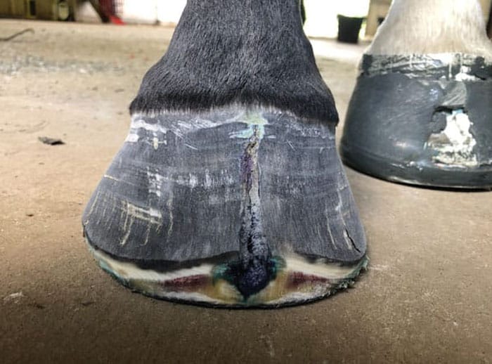
Third Shoeing: The coronet band is showing some new growth. The toe crack is partially stabilized after just 4 weeks. The toe crack has been treated with copper sulfate to limit bacterial or fungal infection. Trimming of the foot continues to focus on the origin of distortion. Bruising in the toe region, in this case, has accompanied the dorsal migration of the foot.
Coffin Bone Remodeling
Radiographically, horses that present with thin soles can also display some coffin bone remodeling.9 Typically, coffin bone remodeling will be acquired over long periods of time. This can be diagnosed through a radiograph from a dorsal-ventral view. The mediolateral distal borders of the coffin bone may show some asymmetry in horses with compromised feet. In the author’s opinion, this often corresponds to conformational deviations in the affected limb. If significant osteolysis is present, careful attention to interpreting mediolateral hoof balance via radiograph may be needed as the distal borders and shape of the coffin bone may not be a reliable reference for trimming.
If the hoof capsule has endured prolonged periods of locomotive stress or the inability to functionally reduce the vibrations of movement, the stresses that aren’t dissipated by the wall structure and lamellar bed could be redirected toward the pedal bone structure.1 Bony inflammation in this region is termed “pedal osteitis.” Pedal osteitis is commonly associated when the vascular channels found near the solar margin of the coffin bone are widened.
Radiographically, pedal osteitis can be present in one or more feet. Commonly, the distal border of the pedal bone will appear somewhat translucent and the margins of the pedal bone will appear inflamed or stressed. There can be multiple presentations of the condition and a lot of variance in the lameness that accompanies the acute, pathological change. While bony inflammation is considered to have a possible correlation to lameness, the underlying origin of the condition should be considered. For some horses, this can be a compounding scenario with other links to the inflammatory condition.10
The pedal bone’s adaption to loading force and inflammation could also be associated with palmar/plantar angle orientation. Horses that are diagnosed with pedal osteitis and high or low palmar or plantar angles, often present with reduced bone density in their respective region of the foot where the bone is showing the bony change. In this context, there is a variance of “normal” with pedal osteitis and the underlying, primary condition needs to be evaluated in regard to the onset of the inflammation and quality of the foot.10
The reduction in bone quality at the distal margin of the pedal bone, which is due to inflammation, has also been linked to rim fractures of the distal phalanx and/or sepsis of the pedal bone.11 Septic pedal osteitis is often treated with surgical bone debridement or sterile maggot therapy in clinical cases.11 A bone sequestrum can be an interesting case for both veterinarians and farriers to work on. Both professions will bring different strengths to the case. The farrier’s role often focuses on offering support and protection, while the veterinarian often offers systemic support.
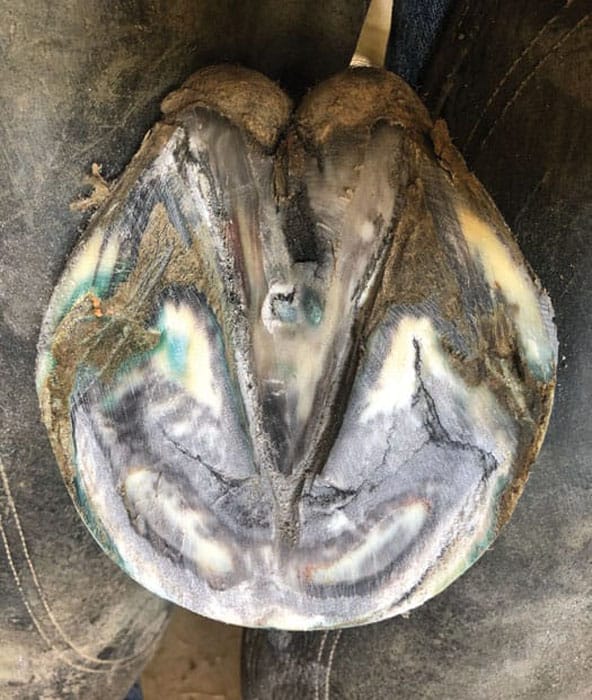
Third Shoeing: The sole continues to perfuse better quality foot. This adds structural integrity to the whole hoof capsule. The heel structure is starting to allow more plantar loading.
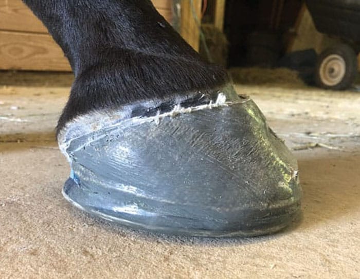
Lateral view 3 months after the initial shoeing.
Therapeutic Support
In cases where the hoof capsule isn’t performing to the optimum level and where there is internal pathology, careful consideration needs to be taken toward the short- and long-term goals for the case. The application of therapeutic support can bring significant value and comfort to the horse. However, without considering the primary onset, the condition may continue to arise if the primary cause isn’t identified.
The hoof capsule is a complex structure and all the anatomical and physiological components must work in unity with each other to offer the horse the best chance at achieving biomechanical efficiency. While it can be easy to be hyper-fixated on certain components of the horse’s distal limb, the health of all the external structures plays a vital role in the overall well-being of the internal structures.
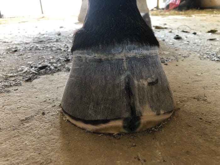
6 months after the initial shoeing and evaluation. The wall structure is improving. The toe crack is stable and no longer needs the aluminum band glued on. The hairline at the proximal margin of the crack has settled.
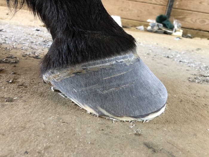
Lateral view at 6 months post the initial evaluation. Trimming is still focused on creating a better foot with improvements in balance proportions.
This case has still got a long way to go. The horse is sound and has begun light riding. The value of the shoeing therapy will slowly be reduced as the horse continues to improve.
Attempting to aid the hoof capsule’s normal characteristics to influence healthy hoof capsule performance can be a valuable asset to bring to each shoeing case. Horses that present with poor hoof quality may need a completely different shoeing package over a horse that is growing better foot quality but has a conformational fault that impacts the horse’s ability to dissipate concussion in a different region of the foot.
Hoof balance is a vital component of good foot care and remaining pragmatic about foot health and function is also of importance. As hoof-care providers, when we evaluate the functionality of our workmanship, the horse’s foot performance can give us valuable information as to whether we may need to apply something that allows more or less therapeutic value.
With all this in mind, it can make equine foot care extensively challenging. The smallest sign of change may indicate the horse modified its gait pattern, or possibly that the horse is finding comfort around a lameness. Therefore, it can also make foot care very fluid, and as foot-care providers, it is our job to monitor the changes and implement well thought-out work responsibly.
References
- Morrison S. (2013) The Thoroughbred Racehorse Foot: Evaluation and Management of Common Problems. Lexington, Ky.: Rood and Riddle Equine Hospital.
- Redden R. Using the Radiographic “Platinum” Standard to Improve Footcare. International Equine Veterinarian March/April 2013 edition.
- Ramey P. (2011) Care and Rehabilitation of the Equine Foot. Georgia: Hoof Rehabilitation Publishing LLC.
- Peterson M, Roepstorff L, Thomason J, Mahaffey C, McIlwraith CW. United States: Grayson Jockey Club. Racing Surfaces, White Paper.
- Curtis S. (2006) Corrective Farriery: A textbook of remedial horseshoeing, Volume 2. Newmarket, Suffolk, UK: R&W Communications.
- Bowker R. (2003) Contrasting Structural Morphologies of “Good” and “Bad” Footed Horses. East Lansing, Mich: Department of Pathology and Diagnostic Investigation, Michigan State University.
- Hickman J, Humphrey M. (1988) Hickman’s Farriery: Second Edition. London, Great Britain: J.A. Allen & Co. Ltd.
- Muir S. (2020) How to shoe for quarter cracks. In: AAEP Virtual Conference Proceedings.
- Fischer MD, Fisher SL. Bone Remodeling of the Equine Distal Limb.
- Dyson S. Nonseptic osteitis of the distal phalanx and its palmar processes.
- Cauvin ERJ, Munroe GA. Septic Osteitis of the Distal Phalanx: findings and surgical treatment in 18 cases.




Post a comment
Report Abusive Comment