Farrier Takeaways
- One effective method of increasing your understanding of the equine anatomy that underlies the conformation of the horse’s hindquarter is drawing.
- The conformation of a horse’s hind limb can accurately be assessed only by means of a plane that bisects the limb.
- The horse’s living limb is structured by joints that are cantilevered in three dimensions and functions by a complex, subtle spiraling in-and-out.
“The anatomist has to observe….to picture…the body not as a surface but as a [three-dimensional] space, in order to understand which he must in his imagination walk through the anatomical elements and perceive what lies behind them.”1
My first task in beginning this series on the anatomy and function of the equine hind limb is to teach the anatomy that underlies the conformation of the horse’s hindquarter.
There is no better way to learn anatomy than to enroll in a class where drawing is required. I have participated in and taught such classes, both for university students and general public.
A hundred years ago, this was the way anatomy was taught, and whether the students were hopeful of becoming veterinarians, medical doctors, anatomy professors or paleontologists, they were required to turn in a notebook of their drawings before receiving a passing grade.
Let me add from the outset: I don’t want to hear anyone exclaim “but I can’t draw,” because all normal human beings can draw.2 In any exercises I might suggest, the expectation is just that you give your best effort; an honest try will yield rewards that you may not have foreseen. Drawing or modeling in clay is so powerful that it will change the structure of your brain.2,3
The Farrier as Artist
In the January/February 2020 issue of American Farriers Journal, I noticed “Recognize Distortion by Drawing the Hoof” by Chris Niclas. This article is intended to guide farriers through drawing the hoof — a great idea, and here’s why.
“Anatomy and art share so many features in common that it is not an overstatement to say that an anatomist needs art,” writes Alexandro Mavrodi and colleagues in The Italian Journal of Anatomy and Embryology.1 “The ability to draw is an irreplaceable qualification [because] when the … student attempts to depict the relations among the anatomical elements and overcome the trouble he faces in this effort, he is not only forced to more deeply understand his science, but he also discovers important details. Exactly as the study of anatomy disciplines the eye of the artist, the creation of anatomical models disciplines the doctor’s hand and especially the surgeon’s,” — and, I would add, the farrier’s.
Judging Conformation the Right Way
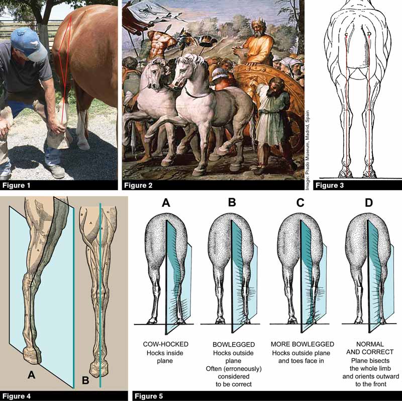
Figure 1. Farrier Sean Finn sets my gelding Oliver’s hoof on his knee to demonstrate that when a normal, correctly conformed horse lifts its hind hoof or swings the hindlimb forward, the midline plane of the limb orients outward toward the front so that the stifle will clear the last rib.
Figure 2. Renaissance artist Rafael painted “The Triumph of King David” in 1517, demonstrating a correct understanding of hindlimb functioning.
Figure 3. Gobaux and Barrier’s 1893 illustration of a horse’s conformation and stance as seen from the rear is a mere diagram; no correctly conformed horse stands with the midline plane of the stifles facing outward (diverging from the midline plane of the body) while the midline plane of the hocks, cannon bones and hoofs faces straight forward (parallel to the midline plane of the body). The horse’s hind limb cannot accurately or correctly be judged by “dropping a plumb line” from the points of buttock down to the hoofs — this must be done by means of a plane, not a line. This illustration has been highly influential over the last century and extremely destructive. It has been reproduced or copied by other artists thousands of times since 1893 and is in a major way responsible for championships being awarded to incorrectly conformed horses and for breeders seeking to produce horses with the twisted structure and stance that it portrays.
Figure 4. The ancients knew that the “plumb line” dropped down the back of the hind limb in View B is only the leading edge of an imaginary plane (seen bisecting the limb in oblique View A (see also other illustrations in this article from Carlo Ruini’s 1598 treatise, Anatomia del Cavallo).
Figure 5. The correct way to evaluate the conformation of a horse’s hind limb in rear view (illustration by the author).
Innovations in Anatomy Study
The history of anatomy has been re-viewed many times;4 the following is but a skeleton outline. The first Greek anatomists were Alcmaeon (500 BCE), Empedocles (ca. 450 BCE), and Hippocrates (ca. 400 BCE), who is called “the father of medicine.” Aristotle (ca. 300 BCE) dissected animals and is the founder of comparative anatomy and also embryology (he studied chick embryos).
Herophilus of Chalcedon (in ancient western Turkey) lived at the same time as Aristotle and is credited with the first known human dissection. He also began the practice of assigning a definite name to each anatomical structure.
Claudius Galenus (“Galen,” ca. 200 CE), a Greek physician who worked in Rome, is regarded as one of the most important early authorities on anatomy.
“The dissection of the animal,” taught Galen, “will teach the seat, the number, the peculiar matter, the size, the shape, and the composition of every part of the body.”
After the end of the Roman Empire in 417 CE, there ensued a period of over 800 years — the so-called “Dark Ages” — when observational science was not generally practiced or was even forbidden in countries ruled by Christians. During this period, the light of systematic observation and scientific thought survived in Jewish communities5 and in countries ruled by Islam.6 The European Renaissance brought about a significant freeing of thought, resulting in a virtual explosion of knowledge of the natural world, including horse anatomy and conformation.
Michelangelo, Donatello, Raphael and Leonardo Da Vinci — all of whom flourished in the late 14th and early 15th centuries — led a large field of lesser-known Italian artists in practicing dissection of horses and other animals. Leonardo Da Vinci produced hundreds of anatomical drawings, and his efforts to understand how to realistically draw horses in rear view are directly relevant here (Figure 14).
Andreas Vesalius (1514-1564 CE) was born in Flanders but worked in Italy and Spain. He wrote one of the great foundational books of medical science, De Humani Corporis Fabrica (On the Makeup of the Human Body). This encyclopedic book represents the first major collaboration between an anatomist and a professional artist (Vesalius drew many illustrations himself, but worked with Jan Calcar, a student of the painter Titian, and other artists).
Vesalius introduced the concept of anatomy instruction that we still use today: observation in the dissecting room aided and reinforced by study of detailed illustrations. Moveable-type printing presses became common in Europe in Vesalius’ time, an important development as they made illustrating a book much easier and helped to spread the concept of the illustrated anatomy textbook across Europe.
It is a gross error to believe that because a work is old it must be of little scientific value. Carlo Ruini (1530-1598) was the most noted horse anatomist of the Renaissance. His illustrations of the horse’s hindquarter and hind limb bones are reproduced here for your enjoyment and study because in all major details, they are correct despite being over 400 years old (Figures 4, 6 and 15).
The Spiral Structure of the Hind Limb
In Figure 6 we see a drawing of bones of the hind limb, from Carlo Ruini’s Anatomia del Cavallo published 423 years ago. The drawings are accurate and clear. I invite readers to compare the same bones and views as presented in any modern horse anatomy book.
Roman numeral I shows bones of the left hind limb in lateral view. C in this view is the head of the femur. H = patella; O = fibula. N = fibular tarsal (calcaneus); I = tibial tarsal (astragalus). M = central tarsal; X = 3rd tarsal. F = metatarsal II (medial splint bone); Q = sesamoid; R = 1st and 2nd phalanges (long and short pastern bones); RT = coffin bone.
II: Bones of the right hind limb in lateral view. In this view, A is the head of the femur, O = central tarsal and E = 3rd tarsal. P = Metatarsal IV (lateral splint bone). View V shows the right patella (R) and patellar cartilage (Q).
III and IIII: Posterior and anterior views of the left femur, in perspective with the distal end closer to the viewer. A = head, E = major trochanter, G = condyles, H = trochlea, I = epicondyles.
VI and VII: antero-lateral and anterior views of the left tibia. A, B = articular surfaces; D’ = spine; B = tuberosity; C = fibula; D = crest. View VIII shows the tibia in perspective with the distal end closer to the viewer. P = articular grooves. The popliteal notch is between A and B.
XI, XIII: Posterior and anterior views of metatarsal bones (cannon bone and splints). A = articular surface, B and C = metatarsal III (cannon bone); P = metatarsals II and IV (splint bones). D = distal articular surfaces; O = sagittal ridge. Note that the specimen is from a young horse, probably less than four years old, as the line of fusion between the shaft and distal epiphysis shows clearly.
IX, X, XI: Views of the fibular and tibial tarsal bones (calcaneum and astragalus). N = tuber calcis; I = trochlear ridges; F = distal tuberosity of tibial tarsal bone. P, S, T = facets for articulation of fibular with tibial tarsal.
XIIII: Distal phalanx (coffin bone) in plantar view. A = plantar surface, B = articular surface.
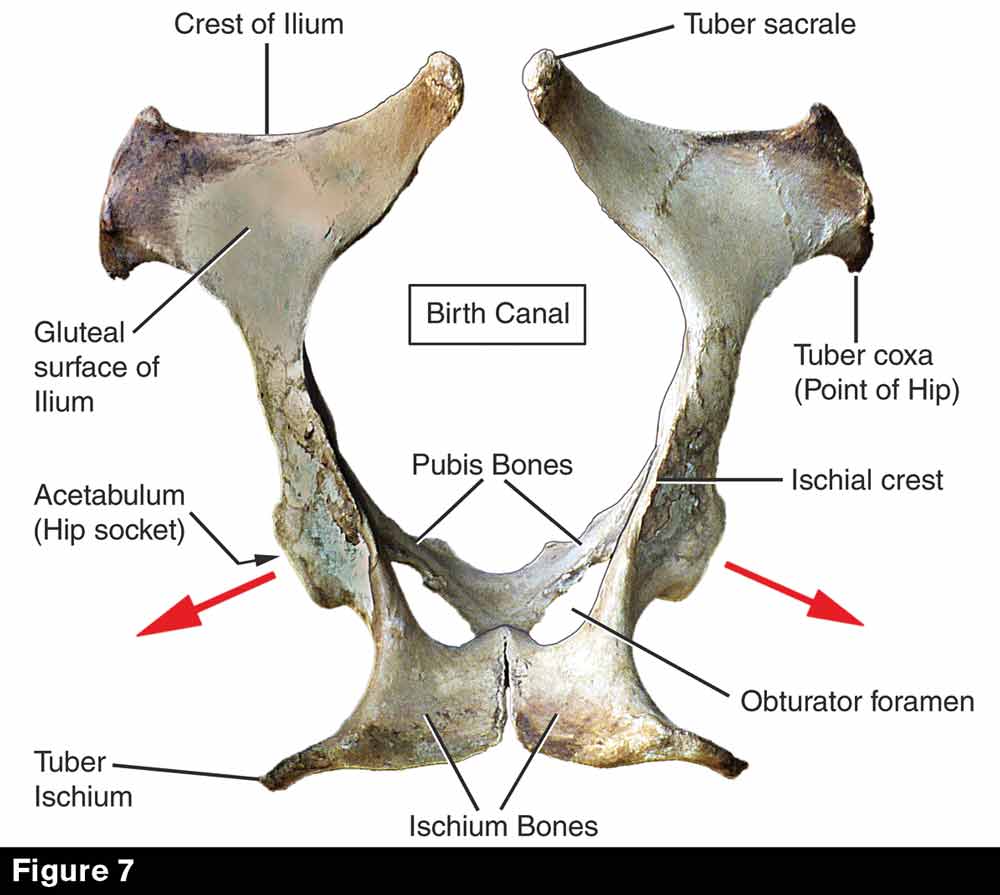
Figure 7. Equine pelvis in dorsal view (i.e. as seen from above); anterior to top. Red arrows indicate the orientation of the top of the plane of fore-aft motion of the femur, i.e. the axis around which it swings.
Figure 8. Closeup study of the bones of the equine coxofemoral (hip) joint. The red line shows the top of the midline plane of the shaft and distal end of the femur. The blue arrows show that the head of the femur fits into the acetabulum (hip socket) at an angle to the midline plane of the body. In total (red and blue planes) this causes the femur to protract (swing forward) at an angle wide of the midline plane of the body, guaranteeing that the stifle joint will clear the last rib.
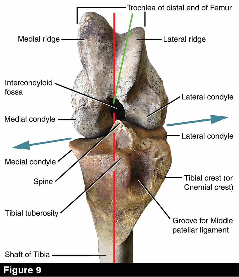
Figure 9. Closeup study of the bones of the equine femoro-tibial (stifle) joint, viewed from directly in front. The patella has been removed. The joint is flexed wide open, with the tibia vertical and the shaft of the femur running back 90 degrees into the page. Red line marks the edge of the plane that would bisect the shaft of the tibia; green line marks the midline plane of the shaft of the femur. The joint folds in the red plane, which is at an angle to the midline plane of the femur. Blue arrows show that the plane of articulation between the articular condyles of the femur and the articular surfaces at the top of the tibia is also tilted (see Figures 1, 8, and 14).
Figure 10. Bones of the equine tibiotarsal (hock) joint. View A, distal end of the tibia; B, anterior face of the hock and cannon bones. Red line marks the edge of the midline plane of the tibia and cannon bones. Purple line shows the orientation of the shaft of the calcaneum, at an angle to the midline plane of the cannon bone. Blue lines show oblique orientation of the distal articular grooves and the ridges of the tibial tarsal bone which fit into them. The tongue-and-groove joint so formed is quite deep and strong, forcing the cannon bone (and pasterns and hoof) to orient outward with respect to the midline plane of the body when the hock is flexed. Note that the obliquity of this joint is coordinate with the orientation of the tibia under the femur, so that when the hock joint is flexed the midline plane of the cannon bone (and pasterns and hoof) aligns only a little bit outward from the midline plane of the tibia (see Figure 1).
After Ruini, a group of Dutch, German, Italian, French and English anatomists bring us up to the 1766 publication of George Stubbs’ The Anatomy of the Horse. Stubbs’ work — both dissections and paintings — is of the highest quality; he is considered the first modern horse anatomist. Sixty-five years after Stubbs, the French anatomist Jean-Baptiste Marc Bourgery in conjunction with illustrator Nicolas Henri-Jacob produced Complete Treatise on Human Anatomy (published 1831-1834), a huge work containing 709 plates with 3,750 individual figures. This book is important not only because of its size and completeness but because it is a masterpiece of anatomical illustration. The authors took advantage of the new process of lithography, which allowed for much more accurate and detailed drawings than the Renaissance woodcuts of Ruini or even the 18th century copperplate engravings of Stubbs.
Building upon the advent of lithography comes at last a name familiar to modern anatomy students: Henry Gray. Gray teamed with illustrator Henry Vandyke Carter to produce the first (1859) edition of Anatomy of the Human Body — later shortened to “Gray’s Anatomy.” Thirty-nine years later in 1898, the German team of Wilhelm Ellenberger and Hermann Baum (anatomists) and Hermann Dittrich (scientific illustrator) produced Anatomie für Kunstlers (Anatomy for Artists), which included a complete set of lucid and exquisitely detailed plates covering the anatomy of domesticated horses, cattle, dogs, sheep, goats, lions and deer.
The plates relating to the domesticated species underwent a “migration” into another extremely important book — The Anatomy of the Domestic Animals — originally authored by Septimus Sisson, a British veterinary student who came to America in the 1880s.
Sisson, who had studied anatomy at two German universities and spoke fluent German, was hired to translate Ellenberger and Baum’s text into English — and Dittrich’s illustrations were naturally reprinted with this edition.
After getting his DVS and being hired to teach anatomy at the College of Veterinary Medicine at Ohio State University, Sisson re-wrote the text to make it more relevant and useful to veterinary students and — naturally — made use of the same set of illustrations, whose superb quality has never been equaled.
In 1901, veterinarian James Grossman, who had been Sisson’s student, revised the text, packaged it with the same set of illustrations, and produced the first edition of the all-time classic Sisson and Grossman’s Anatomy of the Domestic Animals. Today, “borrowing” Dittrich’s plates in this way would be highly illegal, but international copyright law had not been established at that time — and the plates have long passed into the public domain.
The Anatomy of the Domestic Animals has recently been revised again (by James Getty, who had been a student of both Sisson and Grossman) and the book (now a two-volume set) is in wide use.
The history of horse anatomy study would not be complete without mentioning the contributions of vertebrate paleontologists. These are men of my own academic discipline and heroes of my student days: Edward Drinker Cope, Othniel C. Marsh, Henry Fairfield Osborn, Joseph Leidy and William Berryman Scott. Writing between about 1875 and 1920, all of them worked with highly skilled illustrator-draftsmen and their books and technical papers are filled with anatomically accurate, highly detailed illustrations which are also beautiful to look at. Particularly relevant is Osborn’s 1911 Iconographic Type Revision of the North American Equidae, which shows the detailed osteology of all the fossil horses which had been recovered from North American strata up to that time.
Correcting a Destructive Error
Well-known equestrian artist Sam Savitt once observed that a drawing of a horse “is a kind of test … showing how much knowledge of horses the artist actually has.” Artistic representations of horses in both sculpture and painting, no matter what their era and no matter what their style so long as that be reasonably realistic, reveal at first glance what the artist knew. I reproduce several famous equestrian sculptures and drawings here that prove that artists living long ago often had a better grasp of equine hind limb anatomy and functioning than do modern conformation manuals, breed associations and horse show judges.
As Betty Edwards documents in her seminal work Drawing on the Right Side of the Brain,2 when adults are asked to draw (for example) an image of their own hand, what they commonly produce as a first effort is not an image of a hand as it really looks, but instead a diagram — a circle with five sticks radiating outward. People who have spent years reading and ciphering have exercised primarily the left hemisphere of the brain that specializes in organization, classification, tabulation and diagrammatic representation. What initially occurs in art class is that this well-oiled circuitry simply takes over, blocking out any contribution that the right hemisphere might have tried to make in the way of nuanced detail.
Conformation judging — including assessment of the horse’s limbs and stance made by farriers — is plagued by errors arising from this same mental phenomenon.
An illustration of the horse’s hindquarter in rear view that appears in Armand Gobaux and Gustav Barrier’s important 1893 work The Exterior of the Horse is a case in point: no correctly structured horse ever stands in the manner shown (Figure 3).
The illustration treats the horse’s hind limb diagrammatically — as if it were the leg of a table. In fact, the horse’s living limb is structured by joints that are cantilevered in three dimensions and functions not by swinging back and forth like a broom handle, but by a complex, subtle spiraling in-and-out. Gobaux and Barrier’s illustration falsely gives the impression that the equine hind limb is so simple that it can be described, characterized, and analyzed by means of a plumb line. In reality, the conformation of a horse’s hind limb can accurately be assessed only by means of a plane that bisects the limb. A “plane of assessment” must be used to differentiate correct, normal structure and stance from cow hocks and bowleggedness (Figure 5).
The figures presented in this article challenge you to take a careful look at the reality of equine hind limb conformation. I encourage you to take your own photos of various horses so that you can learn to correctly assess hindlimb conformation by visualizing two planes: the midline plane that splits the horse’s body into left and right halves, and the planes that bisect the individual hind limbs. Closeup bone studies presented here show that the horse’s hip, stifle and hock joints are tilted and asymmetrical so that they cannot open or close parallel to the midline of the body.
What did the Artist Know?
Does the farrier know as much about horses as the artist? Look at the hind limb in each of these examples and visualize the midline plane of the limb with respect to the midline plane of the body as a whole.
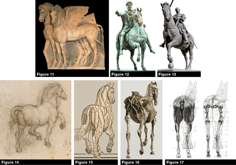
Figure 11. The famous winged horses of Tarquini, in northern Italy, carved by an anonymous Etruscan in about 300 BCE. The artist correctly understood equine hind limb stance, although (possibly because of the limitations of bas-relief as opposed to sculpture in the round) the near hind hoof is twisted inward and thus seen in lateral rather than oblique view. In a well-conformed horse, the toe of the hoof would angle outward so that the plane that bisects the stifle would also bisect the ankle and hoof.
Figure 12. Probably the most famous and influential equestrian statue ever created, this realization of the Roman emperor Marcus Aurelius was made in about 170 CE by a master sculptor whose name is not known to us. Look at the horse’s hind limbs; the artist clearly knew that the midline plane of the horse’s hind limb widens to the front. Note that when the horse’s hind limb is protracted, the amount of outward orientation increases, while with retraction it decreases until at full rearward stretch it has rotated to a position nearly parallel to the midline plane of the body. This “spiraling” or “out and in” effect is driven by the oblique structure of the horse’s hip, stifle, and hock joints
(Figures 6 through 10, above).
Figure 13. This is another very famous equestrian statue, created by Donatello in about 1450. It portrays Italian mercenary general Erasmo of Narni (nicknamed “Gattamelata” or “the candy-coated tiger”) mounted atop a powerful warhorse. The front view shows either that Donatello did not correctly understand equine hindlimb conformation, or else that the famous general’s horse was bowlegged behind, conformation which results when the midline plane of the hind limb parallels the midline plane of the body (refer to Figure 5B).
Figure 14. A Leonardo Da Vinci sketch made about 1480, with overdrawings showing how the great Renaissance genius worked persistently to perfect his understanding of the anatomy, conformation, stance, and movement of the horse seen in rear view. Like the ancient Greeks and Romans, and like other Renaissance artists, Leonardo practiced animal dissection in order to learn anatomical details.
Figure 15. The horse in rear view as pictured in Carlo Ruini’s 1598 treatise on horse anatomy. His concept, although it is 423 years old, is absolutely correct.
Figure 16. The 18th century “Age of Enlightenment” saw George Stubbs’s work “The Anatomy of the Horse” (1766). Stubbs clearly understood that the plane that bisects each hind limb orients outward with respect to the midline plane of the horse’s body. Since femur, tibia, hock and cannon, pasterns, and coffin bone must all fold and unfold in the same plane, to the degree that the stifles face outward when seen from the front, the hocks must face inward when seen from the rear.
Figure 17. Peerless excellence in anatomical illustration —the horse in rear view from Ellenberger, Baum and Dittrich’s Anatomie für Kunstlers as reproduced in Sisson and Grossman’s Anatomy of the Domestic Animals. The authors make no mistake in presenting the real orientation of the bones of the horse’s hind limb: the midline plane of each hind limb angles outward with respect to the midline plane of the body.
References
- Mavrodi, A., G. Paraskevas, and P. Kitsoulis. 2013. “The history and the art of anatomy: a source of inspiration even today.” Italian Journal of Anatomy and Embryology 118(3): 277-276.
- Edwards, B. 2012. Drawing on the Right Side of the Brain, 4th ed. Tarcher/Penguin Books, New York, 320 pp.
- VanSommers, P. 1984. Drawing and Cognition: Descriptive and Experimental Studies of Graphic Production Processes. Cambridge University Press, New York, 284 pp.
- A.O. Malomo, O.E. Idowu and F.C. Osuagwu. 2006. “Lessons from history: human anatomy from the origin to the Renaissance.” International Journal of Morphology 24(1):99-104.
- Fontaine, R. 2011. “Meteorology and zoology in Medieval Hebrew texts,” in G. Freudenthal, ed., Science in Medieval Jewish Cultures. Cambridge University Press, New York, 217-229 pp.
- Turner, H.R. 1995. Science in Medieval Islam: An Illustrated Introduction. Cambridge University Press, New York, 258 pp.
- Bennett, D.K. 1986. Principles of Conformation Analysis, Vols. I-III. Fleet Street Publishing, Gaithersburg MD, 278 pp.








Post a comment
Report Abusive Comment