Scientists at the University of Wisconsin–Madison have created a diagnostic imaging tool that could help prevent equine injuries through early detection and monitoring: a standing helical computed tomography (CT) scanner named Equina.
J. R. Lund, a resident and clinical instructor in diagnostic imaging at the University of Wisconsin-Madison, School of Veterinary Medicine, described how the new technology works when she spoke at the Midwest Equine Podiatry Conference in Arlington, Wis. in late fall. The conference was sponsored by Lodi Veterinary Clinic and Country View Equine Clinic.
“The way I think about standing CT is it’s like a really souped-up X-Ray,” she says. “X-rays are emitted, they pass through the patient and then they are picked up by the detectors. The main difference is we have more detectors in Equina so it is going to cover a larger piece of anatomy.”
Equina is the first CT scanner on the market to vertically scan the lower legs of a standing, sedated horse and also the first dual-purpose standing CT machine. This means it can scan up and down a patient’s legs and move horizontally to scan the head and neck – three areas of the body where CT is advantageous in teasing out anatomical intricacies.
“We scan the horse once and basically we can manipulate that image into giving us any projection we want,” Lund says. “So, if we wanted to look at it in a sagittal or lateral projection we can do that in the same scan as we would look at it for a DP or an axial projection. We don’t have to do multiple scans for multiple projections.”
Safety Benefits
The technology behind Equina is similar to that used for scanning luggage at airports — specifically the machine in which travelers place luggage at TSA checkpoints. This means the level of radiation leakage is low, and safe enough medical that personnel are able to stay in the same room with the horse during a CT scan.
Another benefit to Equina’s technology coming out of the luggage-scanning industry is it can operate in a variety of climates and rugged environments, evidenced by its ability to withstand a kick up to 3,000 pounds and still function, Lund says.
New Findings
Equina fills a longstanding, unmet need in the diagnosis and treatment of a variety of conditions facing horses and other large animals. Already, more than 150 horses ranging in size from a miniature horse to a draft horse have been scanned at UW Veterinary Care, the teaching hospital of the UW–Madison School of Veterinary Medicine (SVM), using the new system. This has led to findings undetectable by earlier methods, including a brain tumor, an orbital tumor behind the eye, and diseases of the feet, teeth and sinuses.
Equina’s creators hope that as the technology becomes more widely adopted, horses exhibiting lameness or other signs of injury can be screened for early signs of a break and treated before the fracture becomes a serious clinical problem.
 |
|
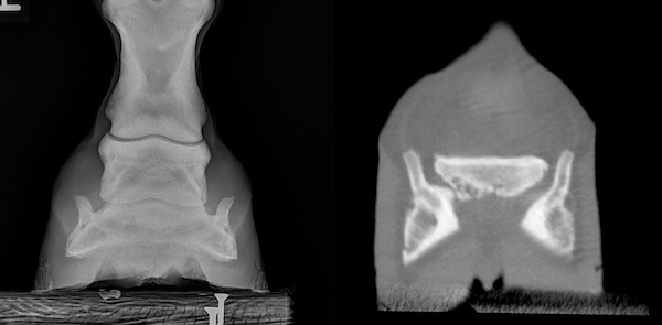
Photo: UW School of Veterinary Medicine A comparison of radiograph, left, and computed tomography (CT), right, imaging of portions of the coffin bone (on the sides) and navicular bone (centrally located) of a horse. Multiple dark regions within the left side of the navicular bone indicate degenerative change consistent with navicular syndrome. CT is much more sensitive to these changes than radiographs and allows clinicians to evaluate the navicular bone in a three-dimensional manner without superimposition. CT also allows for improved evaluation of the soft tissue structures, such as tendons and ligaments. |


