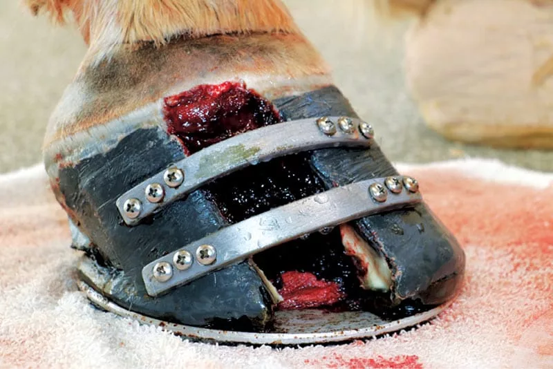
“Avery,” a 12-year-old Suffolk Punch gelding used for trail riding, was referred to the Virginia-Maryland College of Veterinary Medicine (VMCVM) Large Animal Teaching Hospital on June 27, 2018, for further evaluation of a suspected keratoma. Avery had a history of reoccurring sub-solar abscesses and rapidly-progressive lameness on his right thoracic hoof.

Farrier Takeaways

  • There are various options for the surgery team and farrier to approach a keratoma, depending on the case.
  • Your shoeing solution for a keratoma should consider ease of access for the owner in aftercare.
  • Concern for the contralateral shoeing is critical in a keratoma case to help prevent laminitis.

Under physical examination, Avery presented with a 4/5 (American Association of Equine Practitioners grading scale) lameness on his right thoracic limb. He had an increased digital pulse in this foot, while all other digital pulses were within normal limits. Avery was negative to hoof testers in all limbs, except for the affected limb. He had a distinct defect as well as multiple draining tracts in his sole and around the defect of the affected hoof. Paring out this area revealed grey, odoriferous exudate (Figure 1). A palmar digital nerve block was preformed to facilitate probing of the tract.

Following the initial examination, radiographs were obtained. Radiographic examination of the right thoracic hoof and distal phalanx included 0° lateral-medial, 0° dorsopalmar, 65° proximodorsal-palmarodistal oblique, dorsal 65° proximal 45° lateral-palmaromedial oblique, and dorsal 65° proximal 45° medial-palmarolateral oblique. A dorsopalmar 65° oblique radiograph of the left thoracic distal phalanx was acquired for comparison (Figures 2 to 4). The radiographs confirmed a keratoma, given the smooth margins of the defect in the lateral solar margin of the right front distal phalanx with bone resorption and a solar defect secondary to sub-solar abscessing.

file name

On July 2, the abscesses were still draining and the surgery team felt it was best to resolve the abscesses before surgery to decrease the risk of infection. To ensure the abscesses were resolved completely, Avery was put on a 2-week regiment that consisted of daily foot soakings (on days 1 to 3) to flush the draining tracts while keeping the foot bandage wrapped with nitrofurazone to keep the area soft to allow continuing draining (Figure 5). During that time, the healthier sole and frog were treated daily with a betadine solution (days 1 to 5), then switching to 7% tincture iodine to ensure a harder, more sustainable foot for better post-operative support because it would be recruited into load bearing with a urethane support material.a

Discussion of Keratomas

Keratomas are comprised of keratin and can cause focal bone resorption of the distal phalanx, resulting in lameness (Figures 6 and 7). When the bone resorbs due to the mass, the dermal tissue is compromised and the lamellar attachment in that area is nonexistent. The failure of connection between the epidermal lamellae and the bone leaves it susceptible to abscessing. A keratoma is a benign tumor of the keratin-producing cells of the epidermal laminae at the coronary band. The cause is unknown, but it may occur spontaneously or following capsular trauma.

file name

A keratoma causes lameness due to focal pressure on the distal phalanx or the disruption in the connection of dermal and epidermal lamellae, leaving it susceptible for reoccurring abscesses.1 The owner and the clinicians elected to proceed with surgical removal of the keratoma.

A keratoma surgery can be performed standing, with local anesthesia2 that only affects a restricted area of the body or laid down under general anesthesia that affects the whole body with loss of consciousness, with full hoof wall resection3 or removed through a window4 at the location of the mass. The surgery team at VMCVM decided to do the full hoof wall resection to ensure all traces of the keratoma were removed and to further explore the affected area.


The failure of connection between the epidermal lamellae and the bone leaves the foot susceptible to abscessing …


Using radiographic guidance and visual examination, the surgery team used a motorized grinderb with a carbide bitc to score two parallel lines to the non-pigmented stratum medium on either side of the keratoma, as well as a half-circle around the solar aspect of the keratoma marking the resection site. Scoring these lines prior to the surgery helped reduce the time with the Dremel while the horse was under anesthesia. (Figure 8).

Shoeing Following Keratoma Removal

The shoeing principles for a keratoma surgery are:

1

Recruit the frog and as much sole as possible into load sharing to take stress and strain off the lamellar interface.


2

Provide stability to the hoof capsule by inhibiting the inward movement at the top of the hoof wall and outward movement at the bottom of the hoof wall during loading and unloading.


3

Provide a clean environment for the defect post-surgery.


4

Recruit the frog and sole into load sharing to help take strain off of the lamellar interface on the contralateral hoof to help with the prevention of contralateral limb laminitis.


Using these principles to develop a trimming/shoeing plan, the affected hoof was shod with a nailed-on heart-bar shoe made from 17 inches of ½- by 1¼-inch bar stock with a ¼-inch-thick welded-in heart-bar insert with multiple clips (Figure 9). A portion of the shoe was removed to allow access to the keratoma during surgery (Figure 10). A urethane support materiala was applied, leaving access to the keratoma (Figure 11). A hospital plate was made and fit to the shoe utilizing a SoundHorse Series IIIc plated cut out to the size and shape of the shoe (Figure 12). The contralateral hoof was shod with a nailed-on heart-bar shoe made from 16¾ inches of ½ by 1¼-inch bar stock. A urethane support material was applied to the level of the shoe (Figure 13).

file name

After the shoe was applied, two “pillars” made of Poly-Vectrane fabric saturated with a poly-methyl methacrylate adhesivef were placed just outside of each parallel scored line. These “pillars” are designed to be the anchoring point for the screws used to attach two steel bands. One band will be placed proximally to inhibit the inward movement and one distally to inhibit the outward movement.

To make the “pillars,” the Poly-Vectrane fabric was folded into four layers, measured to the vertical length of the hoof wall, then saturated with the adhesivef and applied outside of the lines (Figure 14). The steel bands were forged from pieces of 5/16 x 1-inch mild steel and fit to the contour of the hoof wall. After the shoeing was completed, the hoof was wrapped to keep the foot clean for the surgery the following day.

On the day of the surgery, Avery was anesthetized and put into a left lateral recumbency (Figure 15). The right foot was locally anesthetized with an abaxial sesamoid nerve block. A pneumatic tourniquet was placed on the proximal third metacarpal bone to reduce hemorrhage during the resection.

file name
file name

Using a motorized grinding toolb and a carbide bitc, the scored lines were then extended to the point of capillary oozing and, then using a surgical scalpel, the hoof wall was sharply transected (Figures 16 and 17). Once the lines were cut to the level of sensitive tissue, using pull-offs, the hoof wall/keratoma was pulled upward detaching at the coronary band (Figures 18 to 22). The surgical site was then lavaged and the podiatry team applied the two stabilizing steel bands by screwing into the “pillars” in a transverse plane (Figure 23). The surgical site was then bandaged (Figure 24). The tourniquet was removed, and Avery was sent to recovery.

Post-operatively, Avery was sound at the walk and had normal vital signs. He remained at VMCVM for 1 week post-surgery to ensure proper bandage changes and care (Figure 25). On July 20, Avery was released from VMCVM and scheduled by the podiatry team for a 6-week re-check as well as re-shoeing.

file name

Avery’s Progress

On Aug. 27, Avery was presented to the surgery and podiatry team at VMCVM for a 6-week re-check of the keratoma removal surgery site, as well as to get reshod on the right and left thoracic hoof. Upon arrival, the horse was bright, responsive and sound at the walk. Over the previous weeks, Avery had been on stall rest with occasional hand-walking and grazing. The affected hoof had been receiving bandage changes every other day with application of triple antibiotic ointment to help reduce the risk of infection. After 2 weeks, the surgery site was treated daily with a diluted betadine solution to help with keratinization.

Upon examination (Figures 26 to 29), the surgery site with the once-exposed lamellae had epithelialized and new hoof wall had started growing from the coronary band. During the epithelialization of the lamellae, epidermal basal cells go through mitosis that produce keratinocytes, which then go through a keratinization process. These cells are formed at the basement membrane of the lamella. When they form, they are a complete living cell.

file name
file name

As new cells form through mitosis, the once-complete living cell gets pushed farther away and begins to mature. The nucleus starts to get smaller and the cytoplasm becomes filled with various keratin filaments that form some of the intratubular horn matrix. These cells protect the dermal lamellae by spreading and stacking with one another, eventually covering the whole defect.

The hoof wall goes through a similar, more organized process in re-growing. There are four layers of the hoof wall: there is the stratum inturnum (the epidermal lamellae), non-pigmented stratum medium (the white, less-dense interlayer of the hoof wall), pigmented stratum medium (the sometimes darker, denser outer layer of the hoof wall) and the stratum exturnum (the white waxy layer that is more noticeable just distal of the coronary band). The stratum medium makes up most of the hoof wall, and keratinocytes form in cylindrical patterns called tubules at the coronary band.

LEARN MORE

Watch videos that detail how to approach and treat keratoma cases at
americanfarriers.com/0119

These coronary basal cells undergo mitosis for the remainder of the horses’ life forming the stratum medium of the hoof wall. These cells keratinize like the keratinocyte cells do in the epithelialization of the lamellae.

To guide trimming and shoeing, a radiographic assessment was used to determine hoof growth, medial/lateral balance and dorsal/palmar balance.

For the subsequent shoeing, we decided to reapply a similar set-up as previously described, the nail-on heart-bar shoe with the ¾ Equi-Paka to provide frog and sole support. We then applied the Poly-Vectrane fabric saturated with acrylic adhesive on either side of the defect for the anchoring points for the bands (Figures 30 and 31). The contralateral hoof was shod in a heart-bar with a full Equi-Paka.

Prior to discharge, Avery’s caretakers were directed by the vet team to provide daily iodine treatment utilizing 7% tincture of iodine. The iodine treatment will aid with the keratinization of the tissue, making the defect more dense as well as keeping a more sterile environment. The foot was treated and bandaged, and Avery was scheduled to return for the next recheck and shoeing in 6 weeks.

file name
file name

Learn More

Use Sugar to Treat Those Nasty Hoof Wound Injuries
The remarkable use of sugardine is now accepted more frequently by many veterinarians and farriers because of successful results and its economic significance.


Products

  1. Equi-Pak, Vettec LLC, 600 E. Hueneme Road, Oxnard, CA 93033
  2. Dremel®, Dremel Tool Co., Emerson Electric, 4915 21st, Racine,Wis. 53401
  3. Part 102-17112, C.T.I. Carbide Tools for Industries Inc., 2650 South Grand Ave Santa Ana, Calif. 92705
  4. Series III Plate, SoundHorse Technologies Inc., PO Box 689, Unionville, Pa. 19375
  5. Poly-Vectran, SoundHorse Technologies Inc., PO Box 689, Unionville, Pa. 19375
  6. Equilox®, Equilox International, 110 NE 2nd St., Pine Island, Minn. 55963.

References

  1. Curtis, S. ed., 2002. Corrective farriery: a textbook of remedial horseshoeing. Newmarket Farrier Consultancy.
  2. Jaworska, J. (2018). “Keratoma Can Be Treated Successfully With Simple Hoof Surgery: Hoof resection as a simple way to remove keratoma in a horse.” Journal of the Hellenic Veterinary Medical Society, 67(1), 51-56.
  3. Helms JH, Pleasant RS, Modransky PD. “Management of a Keratoma in a Horse.” Equine Practice, Vol. 16, No. 7, Pg. 35-38.
  4. Getman, L.M., Davidson, E.J., et al., 2011. “Computed tomography or magnetic resonance imaging-
    assisted partial hoof wall resection for keratoma removal.” Veterinary Surgery, 40(6), pp.708-714.


January/February 2019 Issue Contents