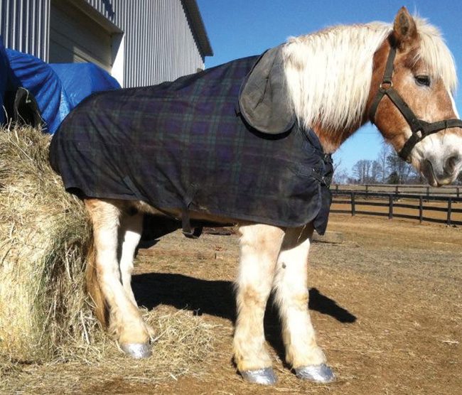
Figure 1. For relief, Bailey developed the habit of leaning or resting his hindquarters on any surface capable of bearing his weight, such as a round hay bale.
A Virginia farrier tackles a case of advanced canker — with favorable results
A 25-year-old Belgian named Bailey was lame, thin and often nipped at his painful feet until they bled when a new owner’s love for the horse brought him to David Giza at Genesis Farriers in Culpepper, Va.
Pain and immobility had resulted in extensive muscular atrophy of the horse’s hindquarters. Knotting and raw sores at the tail head provided evidence that atrophy and hoof pain resulted in Bailey rarely bearing full weight on his hind feet. For relief, he developed the habit of leaning or resting his hindquarters (Figure 1) on any surface capable of bearing his weight, including his new-found farrier.
Bailey had extensive damage to all four hooves, with the right hind being the worst. The issues included extensive flares, infected frogs, swollen heel bulbs, hoof deformities, extensive quarter cracks with protruding growths of proud flesh (Figure 2) and clusters of tentacles hanging from the soles as if he had stepped on an octopus (Figure 3). The crowns of tentacles protruding from the sole easily identified the advanced canker.
Less advanced canker cases share numerous symptoms that are common to a multitude of ailments, so diagnosis should be confirmed with a histopathology/biopsy report.
Canker Conditions
Canker, or equine proliferative pododermatitis, is a rare bacterial hoof disease that is especially common in draft horses. Without immediate care, canker is debilitating.
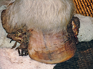
Figure 2. Canker symptoms included extensive flares, infected frogs, swollen heel bulbs, hoof deformities and extensive quarter cracks with protruding growths of proud flesh.
Advanced canker cases often exhibit tumor-like growths, which originate as roots in the live sole. They can develop into thicker stems that grow to the full length of the hoof wall and then extend into crowns of tentacles, which cover the entire sole surface.
The tentacle growth may even protrude beyond the edge of the hoof wall. Although the tentacles have no nerve endings to cause pain, they are vascular, which results in crushing and bleeding of the tentacle tips with every step.
A variety of factors can contribute to the development of canker. The two most directly under the owner’s control are animal husbandry and physical activity.
The primary animal husbandry issue is owner neglect. Whether due to a lack of understanding or commitment, hoof neglect is a near universal factor in equine susceptibility to canker.
Some folks believe that a common precursor to canker seems to be thrush. When left untreated, it can break down the frog, allowing bacterial infiltration and the development of canker.
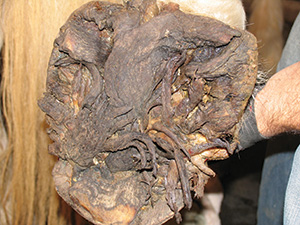
Figure 3. Clusters of tentacles were hanging from the soles.
Another forerunner to canker may be extensive flares, especially with extremely large draft horse hooves. An overgrown hoof wall may result in the impaction of manure or other moist materials that provide ideal conditions for bacterial growth.
A stall-bound or inactive horse is at risk for hoof impaction, while an exercised horse will naturally cleanse the hoof to some extent. Physical activity provides for natural live sole and frog exfoliation as well.
This is not to imply that all owners experiencing canker cases have been neglectful or deficient in their care. Other contributing factors such as animal behavior and environmental conditions are equally important.
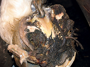
Figure 4. Since the structure of the frog was unidentifiable, it was recreated by carving the collateral sulcus grooves with a hoof knife.
In most cases, controlling environmental conditions such as providing a clean stall and avoiding exposure to wet, low-lying land significantly decreases the likelihood of bacterial growth and the chance of canker development.
Laboratory Diagnosis Essential
In the case of Bailey, a histopathology/biopsy report was obtained from the regional laboratory of Virginia’s Department of Agriculture and Consumer Services. A sample of growth with some live sole attached (approximately 1 inch by 1 inch), was cut from the hoof, sealed in a clean Ziploc bag and sent to the state lab for analysis.
Our veterinarian advised researching a private lab’s expertise with canker tissue samples due to the rarity of canker. Submitting a sample to a private lab, which has little experience with the disease, may result in a misdiagnosis.
Getting Started
The first necessity is a clean, concrete wash stall where extensive hoof cutting and washing can be done. During the beginning stages of treatment, a clean dry stall with deep bedding to provide pressure and help stop the bleeding must be available for the horse to recover after cutting away the canker.
Even during the first days of treatment, physical activity is essential to promote healing. A small dry lot should be provided for light exercise. Food, water and hay should be placed close together so the horse does not have to move very far if in pain. As healing takes place, food, water and hay should be moved farther apart to encourage exercise.
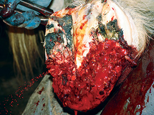
Figure 5. While the tentacles have no nerve endings and are not sensitive, they are vascular and bleed profusely.

Figure 6. Once the tentacles were removed, bactericidal hoof powder was applied and began to congeal with the blood to form a paste.
Hand walking should be done daily on a level surface that has a good footing. As healing progresses, the availability of an indoor arena allows the horse to be worked at liberty and at a trot. Once the horse has fully recovered, it is important that the horse have a job, as a lack of physical activity raises the odds for a recurrence of canker.
Treating The Disease
In Bailey’s case, both a local veterinarian and Dr. Frank K. Reilly of Equine Medical & Surgical Associates in West Chester, Pa., consulted on the case. Dr. Reilly provided a bactericidal hoof powder, which was a critical component to the successful canker treatment.
The six-part canker treatment consisted of an assessment phase, a pre-treatment phase and four canker treatment stages that included:
- Stage 1 (tentacle resection).
- Stage 2 (stem resection).
- Stage 3 (root resection).
- Stage 4 (bactericidal phase).
Assessment Phase
Before beginning treatment, an assessment was necessary to evaluate the overall hoof condition and determine on which hoof to begin. Upon evaluation, Bailey’s most severely affected hoof was the right hind.
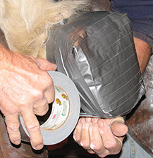
Figure 8. To provide hoof support, durability and cleanliness, a duct-taped boot was applied.
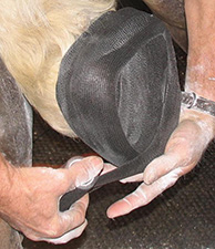
Figure 7. The cotton was secured by wrapping the hoof tightly with Vetrap, which applied direct pressure to control the bleeding.
Clinical signs and symptoms were extensive flare, separation of the toe from the medial and lateral quarters due to extensive quarter cracks extending to the coronary band, protruding proud flesh and canker from the quarter cracks (Figure 2) and canker tentacles in the sole (See Figure 3).
These conditions resulted in an inability to bear weight on the right hind hoof. Since it was determined that the horse would not be able to support himself enough for work to proceed on any of the other three hooves, treatment began with the right hind foot.
Pre-Treatment Phase
The first step in the pre-treatment phase was to clean and assess the hoof. A 4-gallon solution of 50% Listerine and 50% water was prepared and used in an aerated hoof bath to cleanse the hoof. A metal grate placed in the bottom of the bath elevated the hoof to maximize solution penetration between the tentacles.
Once Bailey’s hoof was cleaned and dried, the hoof was trimmed to alleviate stress on the toe and quarter cracks. Tentacles protruding from the live sole blanketed the frog and were brushed aside to find the landmarks for beginning the sole trim.
Even so, the frog and collateral sulcus grooves could not be found. Since the structure of the frog was unidentifiable, it was recreated by carving the collateral sulcus grooves with a hoof knife (Figure 4)before the four canker treatment stages could begin.
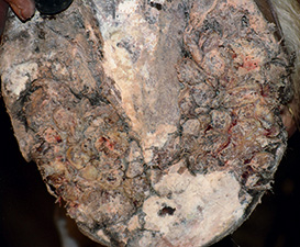
Figure 10. The thicker stem pieces of canker, existing as fleshy growths protruding from the live sole, were removed. A surgical scalpel was used to cut down to live sole, often resulting in heavy bleeding, as seen in Figure 11.
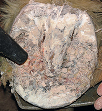
Figure 9. A blower was used to blow out the old powder as attempting to brush it off restarted the bleeding.
Stage 1 — Tentacle Resection
The first step was to remove the crowns of tentacles with hoof nippers. While the tentacles have no nerve endings and are not sensitive, they are vascular and bleed profusely (Figure 5). Once the tentacles were removed, bactericidal hoof powder was applied and began to congeal with the blood to form a paste (Figure 6).
A 2-inch thick layer of cotton was applied on top of the powder to provide cushioning and compression (Figure 6). The cotton was secured by wrapping the hoof tightly with Vetrap, which applied direct pressure to control the bleeding (Figure 7). To provide hoof support, durability, and cleanliness, a duct-taped boot was applied (Figure 8). After the tentacle resection, Bailey was given stall rest for 4 to 6 hours before being given light turnout in the duct-taped boot.
The following week consisted of a daily routine of unwrapping the hoof. A blower was used to blow out the old powder (Figure 9), as attempting to brush off the powder led to reopening the bleeding, reapplying the bactericidal hoof powder and rewrapping the hoof.
Stage 2 — Stem Resection
The next treatment stage began when Bailey began bearing weight on the affected hoof and the canker stems could be touched during evaluation without reopening the bleeding.
This consisted of removing the thicker stem pieces of canker, which existed as fleshy growths protruding from the live sole (Figure 10). A surgical scalpel was used to cut down to the live sole, which often resulted in heavy bleeding.
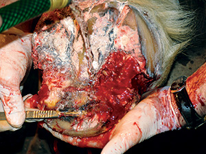
Figure 12. A surgical scalpel was used to cut away at the live sole only those portions containing canker roots and leaving healthy sole untouched.
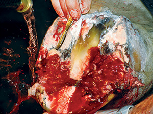
Figure 11. The hoof was irrigated with water to keep the stems visible during resection. The stems were removed in two resections 3 to 5 days apart, as it was imperative not to cut too much or too fast with close proximity to the live sole.
In a prior case, attempts to use a pressure bandage or tourniquet by the attending veterinarian caused the horse to thrash and kick the affected leg. To avoid this concern, the hoof was irrigated with water to keep the stems visible during resection (Figure 11 on Page 66). The stems were removed in two resections 3 to 5 days apart, as it was important not to cut too much or too fast with the close proximity to the live sole.
Protruding proud flesh was removed with the scalpel. Daily powdering and wrapping continued until the powder caused the stems to recede to the level of the live sole.
Stage 3 — Root Resection
The remaining canker was composed of small dry root clusters within the live sole. Giza cautiously cut away at the live sole with a surgical scalpel, removing only those portions containing canker roots and leaving healthy sole untouched (Figure 12).

Figure 13. Bailey appeared to be cured of canker in the right hind hoof at 71 days.
Since removal of live sole can be sensitive or painful, the canker roots were cut with a series of shallow cuts.
At no time was the horse sedated or nerve blocked. It was critical that the horse feel what was happening to the hoof to prevent the possibility of cutting too deeply or causing serious damage.
Whenever the horse began to pull away, it was an indication that the cuts had gone deep enough for that day’s treatment and the bactericidal powder, cotton, Vetrap and duct tape boot were re-applied.
Stage 4 — Bactericidal Phase
Once the canker was removed from the right hind hoof, treatments with the bactericidal hoof powder and wrappings were performed each day. After 7 days, there was no evidence of canker regeneration in the right hind hoof, so the powder and wrapping intervals were extended to 48 hours.
When Bailey was fully bearing weight on the right hind hoof, stage 1 treatment was started on the left front hoof. As each foot was treated and subsequently healed, newly-acquired knowledge was applied to the next hoof until all four hooves were being treated at the same time.
With each consecutive hoof, the bactericidal hoof powder treatments and wraps were continued until removal of the bandage yielded loose powder on dry cotton and no infectious smell, thus indicating that stage 4 of treatment was complete.
At 71 days, Bailey appeared to be cured of canker in his right hind hoof (Figure 13). At 216 days, he appeared canker free in all four hooves (Figure 14).
Post-Procedure & Summation
Like all horses suffering from canker, Bailey will always be at risk for a recurrence of this debilitating disease. With horses recovering from canker, routine hoof care (including regular cleaning and picking) is essential.
The owner or other knowledgeable staffers should closely assess hooves twice a week. The farrier should be notified immediately if there’s any evidence of abnormal growth. The farrier should schedule a full hoof evaluation and trim once a month rather than the normal 6- to 8-week cycle.
As with the diagnosis of canker, a sample of live sole should be submitted to confirm the horse is truly canker free. In Bailey’s case, a sample of live sole was sent to the state lab 10 months after the stage 4 canker treatments were completed. The histopathology/biopsy report confirmed that there was no longer any evidence of canker.
As a result, the now 26-year-old Bailey was officially declared canker free.
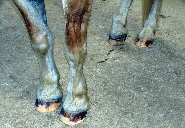
Figure 14. At 216 days, the horse appeared canker free in all four hooves.


Post a comment
Report Abusive Comment