American Farriers Journal
American Farriers Journal is the “hands-on” magazine for professional farriers, equine veterinarians and horse care product and service buyers.
The case presented here is one of many we have been working on. This case is the most interesting because we had the advantage of having a thorough diagnostic history of the horse, which we believe is very important for proper, informed treatment for any patient. This entails having access to quality diagnostic images that pre-date the current treatment protocol.
The following is an example of our approach to treating a certain type of fracture in the navicular bone. Please note that not all navicular bone fractures could be treated this way. Some situations would likely require surgical fixation, but due to the position of the fracture in this case, surgery was avoided.
The case reported here is not an isolated case. We have treated other fractures (e.g. of the pedal bone) with similar methods.
Beau, a 5-year-old, 17-hand, Quarter Horse-Thoroughbred cross, presented with a fracture of the navicular bone of the left front foot on June 2, 2009, at Alamo Pintado Equine Medical Center, Los Olivos, Calif.

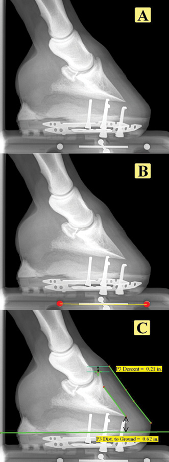
Figure 1: In the original radiograph (A), the metal scale marker built into the block can be seen. In (B) it has been automatically located by the Metron software, which uses it to scale the image so that measurements (C) can be made accurately in the image.
Digital radiographic and magnetic resonance imaging studies were performed to visualize and delineate the extent of the fracture. Surgical fixation was offered to repair the oblique fracture1, but was declined by the client due…