The University of California Davis School of Veterinary Medicine has successfully used positron emission tomography (PET) on a standing horse.
The equine PET software has been pioneered at UC Davis, beginning in 2015. However, the horses being previously imaged had to be put under anesthetic in order to capture the image. Now, horses can remain standing with only slight sedation.
PET imaging technique shows more insight than just radiography or magnetic resonance imaging (MRI). It displays the activity of bone or soft tissue lesions at the molecular level. The equine PET scanner was originally devised from a human brain scanner from Brain Biosciences, Inc. The UC Davis Center for Equine Health (CEH) launched a clinical program using the original human scanner to turn it into a tool to examine horses.
Since 2016, more than 100 horses have been imaged. PET has been particularly useful in discovering bone lesions and distinguishing between active and inactive injuries in the bone or soft tissue. Previous imaging has not been able to provide such in-depth results. However, sedating the horse prevented motion and allowed easy access to the limbs. While this allowed imaging to be done safely and quickly, there were still health risks associated with using anesthesia as well as added costs of having additional staff and equipment.
In developing the equine PET scanner, UC Davis research group led by Dr. Mathieu Spriet joined up with the engineering team from Brain Biosciences, led by David Beylin and Pavel Stepanov. PET images are captured using special detectors arranged in a ring to encompass the horse’s leg. The goal is to create a scanner where the limb is placed within the ring, so the detectors can open freely and also allow the horse to remove its leg without getting hurt on the equipment.
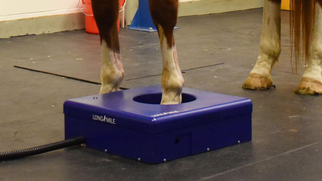
In the meantime, a prototype allows initial validation of standing equine PET. The goal of the final product will mimic that of the human device, but the rings will be horizontal in order to capture the horse’s leg. The diagnostic rings will be encapsulated in a boxy shell to protect the equipment. There is a hole in the center to allow the horse to step inside, but it is not too cumbersome that the horse will have difficulty stepping out of it.
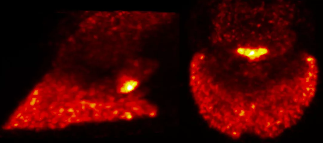
Two horses from the CEH herd were the first to be imaged using the standing equine PET prototype on Jan. 16, 2019. The horses were sedated only as though undergoing a radiograph. The clinician then positioned each horse’s foot in the center of the ring of the scanner. High-quality images were captured within 5 minutes of scanning the foot. Imaging was conducted on 2 consecutive days. Using a bone tracer and a soft tissue tracer, researchers were presented with accurate portrayals of each limb imaged.
Another test is performed by the prototype included the level of radiation the staff was exposed to. The results showed that the staff was exposed to the same levels of radiation that they would for scintigraphic bone scans. These routine scans have proved to be unharmful, which confirms the safety of the equine PET prototype as well.
“This is a major milestone in the development of clinical equine PET imaging,” Spriet says. “The ability to perform PET on standing horses will open many new clinical applications, such as following up on injury healing and screening for lesions at risk for catastrophic breakdown in racehorses.”
More patients should be compatible with this new advent in equine PET scanning now that the need for general anesthesia is no longer required. Training programs won’t be interrupted because the horses imaged won’t need recovery time after the procedure. Laminitic candidates will now receive the benefits of imaging. Previously, this scanning wasn’t available due to the fact that laminitic patients aren’t good anesthesia candidates.
The advent of standing equine PET imaging will allow for a broader clinical population. More than 15 years ago, standing MRI made a major impact on the development of equine imaging. Standing PET had the potential to drastically affect the evaluation of equine lameness.


