American Farriers Journal
American Farriers Journal is the “hands-on” magazine for professional farriers, equine veterinarians and horse care product and service buyers.
Although an uncommon condition, keratoma formation beneath the horse’s hoof wall or sole can cause lameness, recurrent abscesses and damage to the laminae and coffin bone.
Because of the potential for serious medical consequences, keratoma diagnosis, removal and treatment requires either the close collaboration between farrier and veterinarian or the involvement of a veterinarian skilled in farriery.
FARRIER TAKEAWAYS …
The name keratoma implies a keratin-derived tumor, but while the involved tissues are generally mostly keratin, the cells themselves are hyperplastic rather than neoplastic — meaning they have multiplied but aren’t truly cancerous. Keratomas grow slowly over time, but do not spread to other parts of the body.
The keratoma is a spherical or cylindrical mass of keratin that grows between the hoof wall and sensitive laminae or beneath the sole. Lameness develops slowly and sometimes intermittently as the mass begins to press against sensitive structures.
While most keratoma formations are thought to be associated with injury or irritation to the hoof…
American Farriers Journal is the “hands-on” magazine for professional farriers, equine veterinarians and horse care product and service buyers.
American Farriers Journal is the “hands-on” magazine for professional farriers, equine veterinarians and horse care product and service buyers.
Download these helpful knowledge building tools
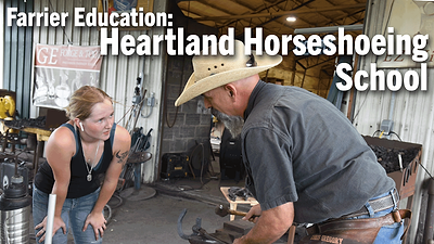
Farriery requires knowledge and skills to provide proper equine hoof care. In this series, sponsored by VICTORY, American Farriers Journal visits Heartland Horseshoeing School in Lamar, Mo. In this edition, Chris Gregory discussed his journey to becoming an educator, his focus in teaching farrier students and the state of farrier education.

