American Farriers Journal
American Farriers Journal is the “hands-on” magazine for professional farriers, equine veterinarians and horse care product and service buyers.
The information, ideas and opinions expressed are those of the author and do not necessarily represent those of the United States Department of Agriculture
Researchers compared to the usefulneness of computed tomography (CT), contract enhanced CT (CECT) and low field magnetic resonance imaging (LFMRI) to identify lesions causing lameness in 31 limbs of 23 horses. All horses had laeness localized to the foot with dagnostic nerve blocks, lameness localized to the foot with diagnostic nerve blocks, and the average duration of lameness was 10 months.
Bone lesions were found in every limb examined with tendon or ligament lesions also identified in 30 of 31 limbs and joint lesions in 28 of 31 limbs. CT identified just two more bone lesions in 28 of 31 limbs. CT identified just two more bone lsions compared with LFMRI, but this was not a statistically significant improvement. Lesions were mot common in the navicular bone, followed by P2 and P3. Lesions of the deep digital flexor tendon (DDFT) were the most common tendon lesions and CT and CECT identified more of these than LFMRI alone. CT failed to identify some DDFT lesions distal to the navicular bone, but identified soft tissue mineralization that MRI did not.
No Single imaging modality proved to be better in every regard. The Authors recommend including evaluation of the pastern when imaging the foot with MRI so lesions that might be clinically relevant are not missed.
-Vallance SA et al. EVJ 2012…
American Farriers Journal is the “hands-on” magazine for professional farriers, equine veterinarians and horse care product and service buyers.
American Farriers Journal is the “hands-on” magazine for professional farriers, equine veterinarians and horse care product and service buyers.
Download these helpful knowledge building tools
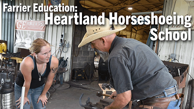
Farriery requires knowledge and skills to provide proper equine hoof care. In this series, sponsored by VICTORY, American Farriers Journal visits Heartland Horseshoeing School in Lamar, Mo. In this edition, Chris Gregory discussed his journey to becoming an educator, his focus in teaching farrier students and the state of farrier education.

