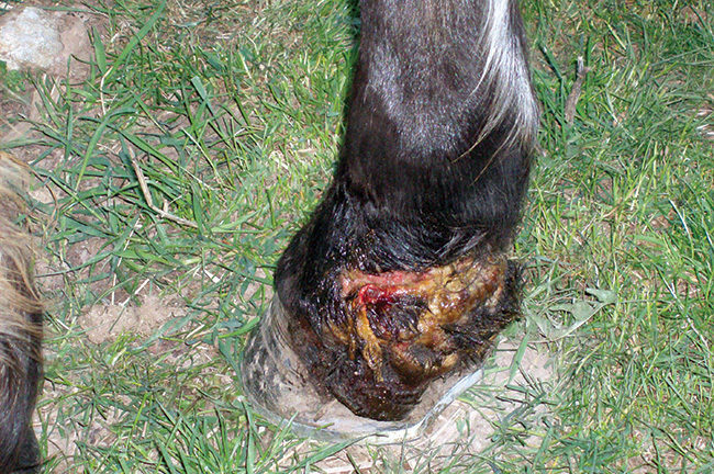
Wire likely sliced through the back of the pastern and into the distal cushion, detaching the back portion of the hoof.
Long-term injury situations often lead to both an emotional and a financial commitment for the horse owner. And it gets serious when tough decisions need to be made. An injury with one of my own horses is an example of how complicated these cases can be.
In 2005, my 1-year-old Quarter Horse gelding came stumbling out of the pasture with a nearly severed hoof. Something, which I assume was wire, had sliced through the back of his pastern into the digital cushion, leaving the back portion of the hoof detached from the leg.
A local vet tried to stitch the broken and torn hoof back together. The vet confirmed my worst fears — Fritz’s injury would likely never heal correctly or maybe not heal at all. With frequent movement, the wound would likely keep reopening, making it impossible to heal.
Daily Care Required
The vet visited every few days for 3 weeks to mend broken stitches, re-wrap the wound and check for infection. Between vet visits, I changed the dressings, cleaned the wounds, applied medicine to flush out infection and yelled at Fritz when he tried to rip off his bandages.
The vet visits dropped to weekly and the wound began to show signs of healing. My daily routine continued for many months. After awhile, the bandage was left off for an occasional day to allow oxygen to reach the wound.
While she wasn’t working on the case at this time, equine veterinarian Julie Bullock says another option for this type of injury would be to cast the leg after treating the infection and taking radiographs.
“The first thing that pops into my brain is casting because you want to get all those structures back together and stop the motion as quickly as possible to ensure the best outcome,” explains the vet from Mount Sidney, Va. “Injuries that involve the hoof capsule and go above the hoof capsule are often dealt with best with casts.”
After 2 years of on-going treatment, Fritz was back in the pasture, but his heels were growing separately from the rest of his hoof due to damage to the coronary band. Hoping to encourage new and solid growth, Brandon White, a farrier from Warm Springs, Va., used a straight bar shoe to relieve pressure and provide heel support.
Sensitive Lamina Infection
All seemed well until the 6-year-old horse was seen limping in the pasture. The sensitive lamina had become infected due to the split in the heels. The sensitive lamina began to swell and push against the hoof wall, causing it to crack. Swelling and infection began to spread toward the knee.
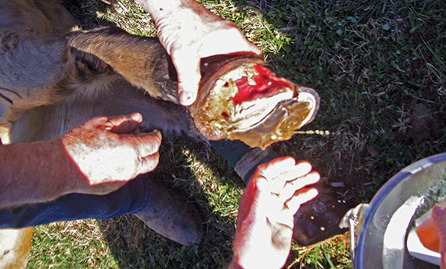
Nearly half of the horse’s damaged hoof wall was removed during surgery.
“I didn’t think there was any hope,” White says. “I’d never seen anything like that.”
The next step was to call Don Cromer of Westwood Animal Hospital in Staunton, Va. “The fracture was through the sensitive lamina all the way from the toe to the cleft of the frog,” the vet explains. “That left the whole outside hoof wall loose and literally hanging from the coronary band.”
Cromer recommended surgery to remove the damaged hoof wall. Even then, hoof regeneration was not a certainty because of unknown damage to the coronary band.
After surgical removal of half the hoof wall, Cromer wrapped the hoof and lower half of the leg heavily and placed the horse in a clean, soft-surfaced stall. A week later, Fritz returned home to a small pen where he spent a year with limited exercise until the hoof re-grew.
Infection Worries
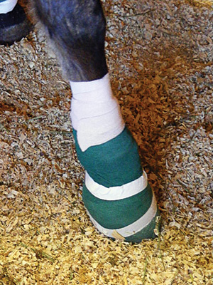
Following surgery, the horse was heavily bandaged with 1 inch of padding that was placed around the hoof in order to provide a solid and stable foundation to stand on while protecting the exposed sensitive lamina.
“I hadn’t seen a hoof that broken away in years,” says Cromer, who felt the biggest task would be keeping infection out of the bone. “So much of it is in the aftercare.”
The vet says it’s important to always keep bandages dry and change dressings as needed. In addition, horses should be kept in a pen with soft ground during the healing process.
“You have to have a place that’s fairly smooth, dry and clean,” says Cromer.
I kept Fritz heavily bandaged with an inch of padding around the hoof to protect the exposed sensitive lamina and provide something solid and stable to stand on. The padding and bandages, which had to be changed every few days, extended up to the knee so extra weight was not attached and supported solely by the weak hoof.
I mixed crushed SMZ antibiotic tablets with molasses-flavored water to help deal with the infection.
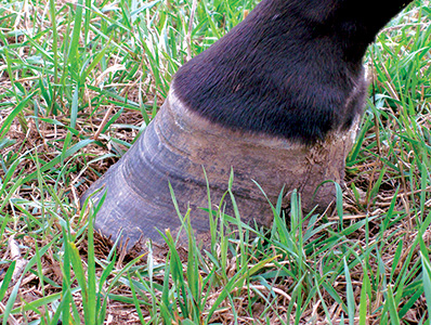
After a month, a hard, white bulge followed the coronary band from the front around the outside of the hoof all the way back to the bulbs. This was regrown hoof, but Fritz still couldn’t place much weight on the injured hoof.
“Once a horse stands on a good leg for an extended period of time it can cause a stress founder,” explains White. “God gave them four legs for a reason.”
White built an acrylic hoof wall so the horse could stand with more weight on the healthy hoof, reducing the risk for stress founder. A shoe was attached to the artificial hoof wall for stabilization. The farrier kept in close contact with the veterinarian throughout the process.
“Any time the hoof is involved and when you need corrective shoes, it’s important that the farrier and vet work together,” says Cromer.
7 Years Of Treatment
After surgery and hoof wall regrowth, there was some rotation in the coffin bone and considerable tension in the deep flexor tendon. A plan was devised by the farrier and veterinarian to relax the deep flexor tendon and relieve extensive pressure on the coffin bone, which was in danger of pushing through the sole.
For the next 9 months, White visited every 4 to 6 weeks to maintain the hoof until he finally removed the shoe and acrylic material to reveal a new hoof wall with connected heels. A straight bar shoe was glued on to prevent further damage from nails.
Fritz was occasionally moved from his soft, dirt-cushioned pen to a small, flat, grassy area that exposed the new hoof wall to harder ground. However, a slight slope leading back to the paddock forced him to limp and sometimes walk on three legs. There was obviously something that still wasn’t quite right.
Bullock offered to take a closer look at the foot. “What we did with Fritz was try to put something on his foot that would be therapeutic, protective and anti-traumatic,” explains Bullock, who decided to glue wooden clogs to the front hooves. “We didn’t have anything to nail to, and I didn’t want to drive nails into the already injured foot.”
After detecting coffin bone rotation, Bullock tried to realign P3 by trimming more off the heels back from the apex of the frog before attaching the clogs.
“Our goal for him is to grow plenty of sole so he’s comfortable and can realign that bone in the hoof capsule,” says Bullock.


Post a comment
Report Abusive Comment