The choices farriers make when trimming and shoeing to achieve a certain result are going to have an impact on the entire equine limb and, at times, may result in unintended consequences. Understanding the anatomy of the equine limb beyond the hoof can help reduce the chances of a farrier’s action having an adverse reaction elsewhere. It can also improve the quality of communication among equine colleagues.
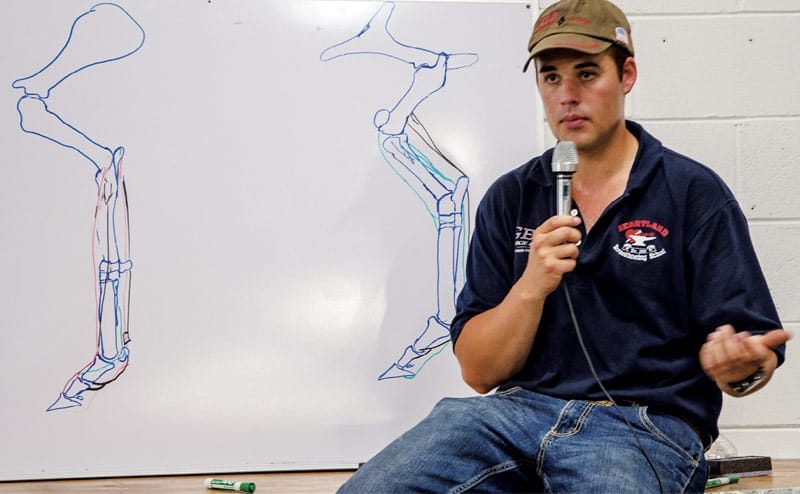
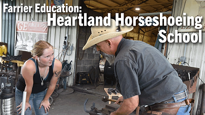
Cody Gregory, an instructor at Heartland Horseshoeing School in Lamar, Mo., offered a primer on equine anatomy as part of the GE Forge and Tool Goodwill Clinic at the grand opening of Ocala’s Farrier Supply. Gregory discussed bones, front and hind limb anatomy, joints and tendons.
Bones in the Front Limb
A horse has five main types of bone structures: flat, sesamoid, long, short and irregular. Each type of bone serves different purposes, Gregory says. Flat bones, like ribs or the skull, protect vital organs. Sesamoids are small bones that allow the horse to change the direction of pull.
Farrier Takeaways
- When discussing the location of bones or other structures within the equine anatomy, using the correct terminology will ensure all members of the horse’s management team are on the same page.
- Raising and changing the angle of the horse’s heel puts more strain on one tendon and less on another — it does not make the weight disappear.
- Improve the accuracy of hind foot trimming by balancing the foot in a weight-bearing position using the tarsus and cannon bone for reference.
“The way that I like to think about a sesamoid is it’s like a crane with cables allowing the crane to lift a bar or anvil,” Gregory says.
Long bones make up the limbs of the horse, giving it height and the ability to propel itself. Short bones, like carpals, provide shock absorption. Irregular bones, like those making up the spinal column, protect the central nervous system.
Bones that make up the front limb, or the thoracic limb, include the scapula, humerus, ulna and radius (which can be recalled using the acronym “SHUR”). Next, is the carpus.
“The carpus is interesting,” Gregory says. “All the flexion of the carpus happens in the proximal two joints on the palmar aspect.”
The proximal row of carpal bones is composed of the radiocarpal bone, the intermediate carpal bone, the ulnar carpal bone and the accessory carpal bone. “That’s the one you’d put your phone in,” Gregory jokes. The distal row is composed of the first through fourth carpal bones.
“When you are talking about numbered anatomy, hold up your hand in front of you, raise your first finger and raise your fourth finger. The first finger is considered medial and the fourth finger is considered lateral. So, applying that logic, C1 and C2 will be medial, C3 will be in the middle and C4 will be on the lateral side.”
Following the carpus are the metacarpals, which include the cannon bone and splint bones. The second metacarpal being the medial splint bone and the fourth metacarpal being the lateral. The large bone between the splint bones is the cannon bone or third metacarpal. This is the largest bone in the horse’s leg, bearing in mind that “leg” is defined as from the knee or hock down. Located at the distal end of the cannon bone is the proximal sesamoids. The palmar surface is smooth, allowing tendons to pass over it.
“Think about what the sesamoids do,” Gregory says. “Whenever the flexor muscles attached to those two tendons on the palmar aspect of the leg contract, they are going to pull and have more leverage and more strength because of those sesamoids. It changes the direction of pull.”
Whatever we do to the foot will have an instant effect on the coffin bone ...
After the proximal sesamoids are the phalanges, numbered 1 through 3. P3 is known as a distal phalanx or coffin bone. On its palmar border is the navicular bone. Like the proximal sesamoids, the navicular helps change the direction of pull.
The main structure that connects the front limb to the horse is the serratus ventralis. The serratus ventralis fans out and creates the thoracic sling, which is a big part of the stay apparatus — the structure enabling the horse to stand with minimal muscular effort.
Joints to Know
The primary joints that farriers deal with are synovial joints, which allow horses to move with ease. A synovial joint is comprised of a capsule that contains an inner lining called the synovial membrane, which secretes a fluid that lubricates the joints. There are three main synovial joints that farriers need to be aware of, Gregory says. The shape of these joints determines how the horse can move. These are: ball-and-socket, plane and hinge.
The ball-and-socket or enarthrodial joints in which a rounded bone fits into a socket of another bone like a ball in a cup, allow the horse the greatest range of motion, Gregory says. The shoulder in the front limb is one example. The hip joint, in the hind limb, is another. Joint encapsulation is more complete in the hip joint than in the shoulder, however, because the horse propels itself with the hind limb and generates more force through the hip joint.
Plane, or arthrodial, joints have bones with articulating surfaces that are flat or slightly curved, Gregory says. These joints allow for gliding movements. Examples of this type of joint can be found in the tarsometatarsal joint of the tarsus.
“The way that joint works,” Gregory says, “as the horse runs and it stops, the tarsus is going to slide across that third metatarsal. It’s going to move in a gliding motion. That’s going to wear and can result in bone spurs.”
In a hinge or giglymus joint, a bone with a slightly rounded end fits into a hollow end of an adjoining bone. One of the bones moves, while the other remains stationary, similar to the sweeping motion of a door. The fetlock is one example of a hinge joint. It allows flexion and extension but lateral movement is limited.
Bones in the Hind Limb
The hind limb of the horse begins at the pelvis and runs to the coffin bone. Like with the front limb, Gregory offers an acronym, PFPFT, to help recall the names of the larger bones making up this structure: pelvis, femur, patella, fibula and tibia.
Not all bones are easy to identify but the next one is hard to miss.
“If you are looking at a picture of a horse’s anatomy, there is a big bone that sticks out at the back called the calcaneus,” Gregory says, “which is the insertion of the calcaneal tendon group.”
Dorsal to the calcaneus is the talus, which sits on top of the horse’s central tarsal, which is shaped similar to and sits on top of the third tarsal. The most lateral tarsal is T4. T1 and T2 are visible on the medial aspect. Next are the metatarsals. The fourth metatarsal, Gregory says, is the biggest and longest splint bone in the horse anatomy.
Like the front limb, the hind limb also has cannon, sesamoids, navicular and P1-P3 bones.
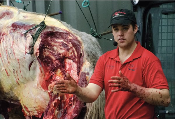
Attending a dissection, like this one led by Cody Gregory at the 2019 American Farrier’s Association Convention, is another way to improve your understanding of equine anatomy.
Tendons and Ligaments
Tendons and ligaments enable proper function and movement of the equine limb. Both fibrous connective tissues, the tendon attaches muscle to bone while the ligament connects bone to bone. How a hoof is balanced will impact how weight is distributed to the flexor tendons.
Tendons are enclosed in synovial sheaths, Gregory says, which enable them to move smoothly. Ligaments also have some elasticity and are for support. Typical tendons are named after the muscle from which they originate.
On the hind limb there are two structures farriers should be familiar with. On the cranial aspect is the peroneus tertius tendon, which originates in the distal end of the femur and inserts into the third metatarsal and fourth tarsal and eventually makes the cunean tendon, Gregory says. On the caudal aspect is the superficial flexor tendon, which originates at the subcondylar fossa on the caudal femur.
These structures make up a unique anatomical feature in the hind limb called the reciprocal apparatus. The definition of the reciprocal apparatus is when the femortibial joint (stifle joint) flexes or extends and the tarsalcrural joint (tarsal metatarsal joint) has to do the same. It’s in part due to the reciprocal apparatus that Gregory recommends evaluating the hind limb balance in a weight bearing position.
“When you bend the tarsus, it pulls that fetlock up,” Gregory says. The fetlock joint only allows movement in two directions. If that cannon bone, that third metatarsal, has any slant on it when you flex, you will cause the fetlock to bend around the cannon bone crookedly. It can tell you a lot of lies.”
Gregory prefers to balance horses by watching how they stand. He looks for a straight line through the tarsus and cannon bone and an even platform beneath it.
Another important tendon is the superficial flexor, which inserts into the calcaneus. The flexor tendons of horses’ lower limbs are important for transferring the muscular energy necessary for speed. The superficial flexor tendon (SFT) is also commonly strained. Proximal to the fetlock joint, the SFT forms a ring-like structure that wraps around the deep flexor tendon (DFT). The protective ring is called the manica flexoria.
LEARN MORE
Enhance your knowledge about equine anatomy by:
- Reading “A Glossary of Therapeutic Farriery Terms.”
- Watching “Anatomy Review: Lower Limb Form and Function.”
Access this content by visiting americanfarriers.com/0919
“If we were to toss a rope over the top of a rafter and I wanted to pull an anvil up on that rope — back and forth, back and forth — what would happen to that rope? It would get frayed. If I put a piece of leather over my rope it would last a lot longer. The manica flexoria is going to have a very similar job.
“When this horse bears weight, the manica flexoria is going to be pulled more into our sesamoid region and it’s going to act as the piece of leather over our sesamoids to protect our deep flexor tendon.”
The SFT bifurcates distal to the fetlock joint and inserts collaterally (both sides of) the distal P1 and proximal P2. Unlike the DFT, it does not continue down into the foot. The primary difference between the thoracic and pelvic limb for the SFT is the insertion at the calcaneus on the hind limb. In the front limb, there is not a similar proximal insertion for the SFT, but there is a check ligament known as the superior check ligament that attaches to the SFT. In the hind limb, there is not a check ligament for the SFT.
The DFT originates from the deep flexor muscle, and courses distad along the medial side of the calcaneus in the hind limb, or medial to the accessory carpal in the front limb, which offers it some extra protection in these areas. It continues distad and inserts in the semi lunar crest of the coffin bone, which is the most distal insertion of any of the tendons in the horse.
A major weight-bearing structure that is located on the palmar/plantar aspect of the leg is the suspensory ligament. The suspensory ligament is responsible for stabilizing the lower limb and providing energy as the horse moves. Injuries to this ligament are common, particularly for equine athletes.
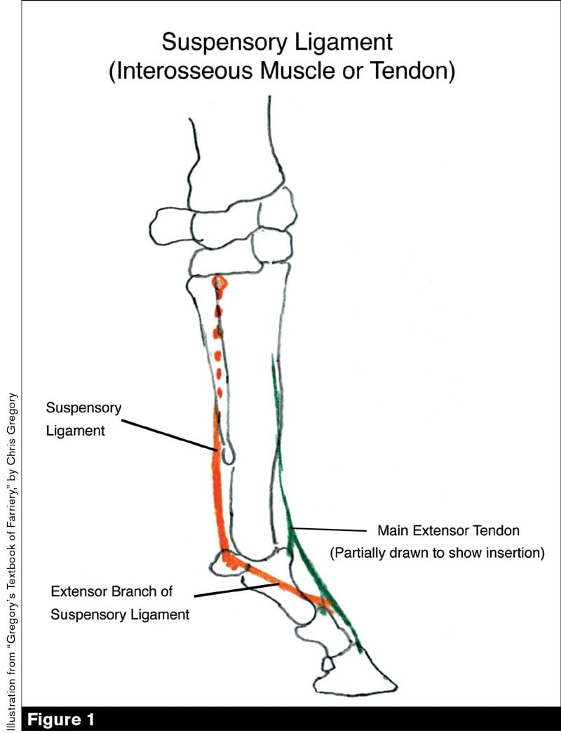
The suspensory ligament (Figure 1) originates at the proximal palmar/plantar cannon bone. It bifurcates at the distal end of the splint bones and inserts into the proximal seasmoids. From that point, it continues with the extensor branches of the suspensory ligament. They course dorso-distad from the sesamoids and insert into the main extensor tendon at about the level of the pastern joint.
There is a lot of misunderstanding about how these tendons work, with some farriers thinking that by raising the horse’s heel they can “take weight off the tendons.”
If a farrier raises the heel, the coffin bone will be steeper as well and the deep flexor tendon will slacken. As a result, the fetlock will descend. When the fetlock descends, the superficial flexor tendon and suspensory ligament become tight.
“Whatever we do to the foot will have an instant effect on our coffin bone,” Gregory says. “You can’t raise the heel to take the weight off of both tendons. By raising and changing the angle of the heels, all we are doing is putting more strain on one tendon and less on the other. You can’t make that weight disappear. That’s hugely important to understand.”
Basic Terminology
When discussing the location of bones or other structures within the equine anatomy, Heartland Horseshoeing School instructor Cody Gregory notes it’s important to use the correct terminology not only to ensure that all the members of the horse’s management team are on the same page, but also to help solidify a farrier’s credibility within the team.
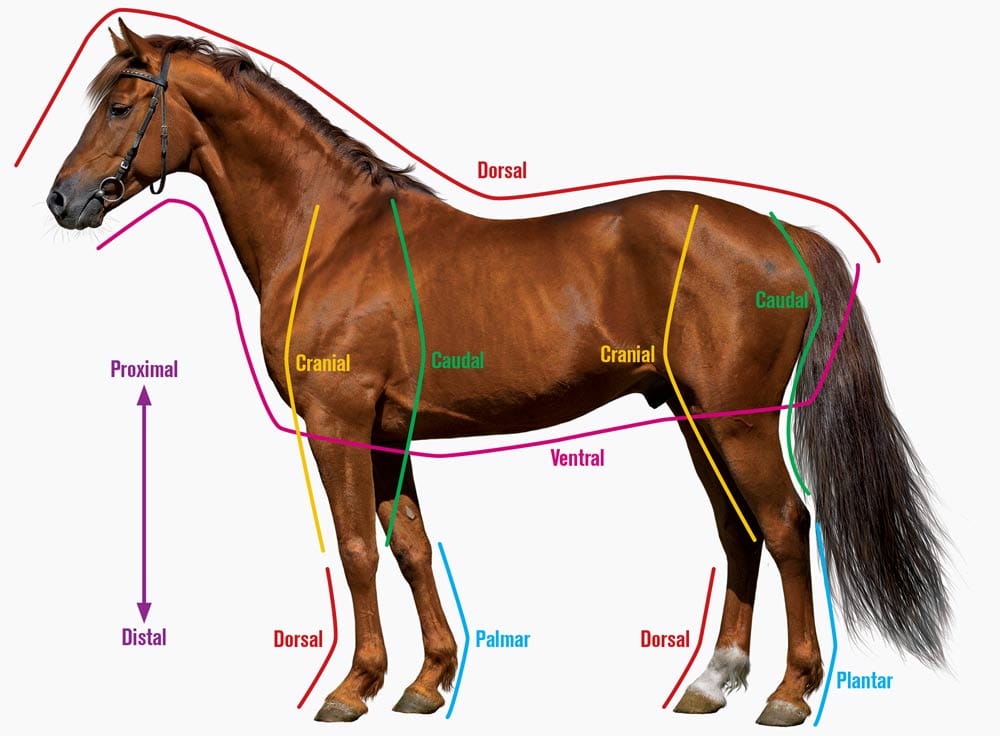
For example, using words like anterior or posterior, which pertain to the front or back of bipeds, would be inaccurate when talking about a horse unless referring to the eyeball, Gregory says. Cranial and caudal are the terms farriers should use.
“Using correct terminology will show equine colleagues you are knowledgeable about what you are talking about,” Gregory says.
“Cranial” is the horse’s structure above the knees and hocks closer to the head, while caudal is the part above the knees and hocks that is closer to the tail.
“Dorsal is going to be toward the front edge below the carpus or tarsus; so toward the head. On the front limb, toward the back on the caudal end of the horse, that would be palmar; on the hind limb, it will be plantar.”
Other important terms to become familiar with are proximal and distal, which mean closer and farther, respectively, relative to center mass. Medial means toward the middle while lateral refers to the outer side.
“You can use incorrect terminology and what you are saying may be understood, but it shows a lack of vocabulary and lack of anatomical study,” Gregory says. “In turn, you will have things explained to you as a layman. If you are a layman, then you are at the mercy of being told to do something and having no way to understand if it is the right or wrong thing to do.”


Post a comment
Report Abusive Comment