In the summer of his 14th year, Lobster showed signs of arthritic changes in both front pastern joints. While his slightly long pasterns make him a candidate for this problem, “high ringbone” was not the anticipated diagnosis. After all, he had been carefully shod and managed his whole life.
It was not a diagnosis I wanted since advanced ringbone — osteoarthritis (OA) of the pastern joint — is painful and progressive. Some form of OA affects the vast majority of performance horses at some point. While OA is incurable, the clinical effects can be treated in an attempt to slow its progression.
Mike Wildenstein, CJF, FWCF (Hons.), the former resident farrier at Cornell University in Ithaca, N.Y., developed a corrective shoeing prescription that took Lobster’s conformation and age into account. Ideas offered by other members of the farrier, vet and the owner team led to new treatment options along with changes in the horse’s management and conditioning program that helped extend Lobster’s career.
In this case history, I’ll describe what I call “aggressive management” of OA in Lobster’s pastern joints. Shoeing and compromise are at the center of this story.
I earlier described Wildenstein’s success at shoeing Lobster’s hind feet to accommodate a base-narrow stance and conformational defects in those limbs (“Education Of A Horse Owner,” American Farriers Journal, September, 2006, Pages 96-100). The farrier’s work and careful explanations convinced me that three key points came into play for managing this ringbone concern:
- Evaluating the hoof and lower limb conformation in the standing horse is only part of the problem. The farrier must also consider the horse in motion and evaluate limb conformation up to the hip or shoulder joint as this will determine the way the horse uses his feet. Wildenstein went further, taking the horse’s environment, management and the rider’s ability into account.
- Farriers do more than solve engineering problems. The biomechanical goal with the front limbs seemed straightforward — raise the heels and correct the angle of the pastern axis from coffin bone to fetlock. But we had to address more than one conformational issue and had to consider the effects of any shoeing strategy in regard to the physiological effects on the internal structures of the foot. In other words, the best engineering solution doesn’t work for all horses at all stages of life.
- The farrier plays a central and distinct role in a team that also includes the rider, trainer, barn manager and veterinarian and owner.
Having invested a great deal of time, money and effort in our performance horses, owners expect a quick solution to any problem.
My experience over the past 3 years suggests an owner’s thinking needs to be changed despite the impressive array of available joint supplements, injections and other treatments. The farrier can provide a uniquely long-term view of OA management.
The Horse’s Contribution
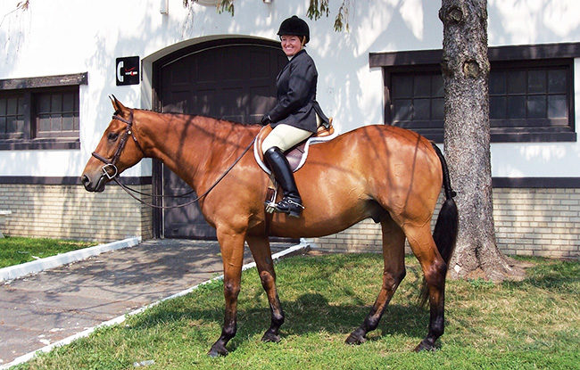
Lobster is a 16.1-hand Dutch warmblood 1,300-pound gelding that was shown in the hunter and equitation divisions. While he has been blessed with generously sized feet, his conformation reflects both the good and bad features of his ancestry. Like most modern performance horses, he was bred to be larger and heavier than his wild ancestors (Figure 1).
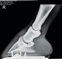
FIGURE 2. In this radiological confirmation of mild ringbone, note the small exostosis at the top of the short pastern bone (P2) and the shallow angle of the coffin bone. This image also reveals fetlock varus conformation.
While none of his conformational defects cause problems by themselves, they do cause parts of the limb to deviate from a single forward/backward plane when the horse is in motion.
The joints between any misshapen bones form the mechanical point where different trajectories of motion are resolved. Since their articulating surfaces do not meet in perfect alignment, the joints’ soft tissues pay the price.
The pastern joint is a particularly unforgiving one and the smallest additions of exostosis, ridges or pearls of bone at their margins comprise the diagnostic signs of OA. In advanced cases, radiographs may reveal disturbance of the subchondral bone near the articulating surfaces, or degeneration within the joint.
Yet by the time radiographs reveal the bony evidence of either type of OA, you’re looking at a long-term pathology.
In well-managed, injury-free horses, the bone remodeling associated with OA simply reflects the consequences of imperfect conformation and physiology for a horse. Since exostoses typically are not reabsorbed even if the offending mechanical issue is resolved, the best we can do is prevent the addition of new bone.
The real problem is irritation and damage to the synovial lining that is responsible for producing healthy joint fluid. As the OA process continues, cartilage at the articulating surfaces of bones is damaged and worn away to the point that inflammation can’t be managed.
Aggressive Diagnosis
While early OA detection is the best defense, I didn’t look for it early enough. Sub-clinical lameness often slips by owners because they see their horses every day.
As a result, it may be up to the farrier to note the small changes in the horse’s stance, behavior, movement and muscling that others miss. Vets tend to see horses only after the owner has identified genuine lameness.
My slow discovery of the arthritic changes may be typical. In the fall of his 13th year, Lobster left the manicured footing of the hunter ring for a couple of hunter paces over uneven footing. While he seemed fit enough for the job, I soon noticed that he began more careful while working his way down hills during our weekly trail rides.
His history and conformation kept me from asking the right questions, and he seemed to be like the horses with diagnosed navicular syndrome that I’d ridden as a kid. But his breeding and large feet did not make him an obvious candidate for degenerating navicular bones.
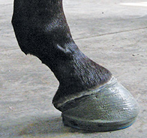
FIGURE 3. Sound Horse Technologies Morrison Roller Motion Series I (blue pad) shoe with a 2-degree polyurethane wedge, right front. This shoe with the softer black pad proved to be the best over-all shoeing strategy. Radiographs revealed the angle at the pastern joint seen through the axis of the bony column was more correct, even though the caudal aspect suggests the pastern joint is broken back or flexed.
Since his suspensory ligaments were a bit sore upon palpation, I assumed the work at speed over varied footing had simply overtaxed the soft tissues high up in the leg. Though I did not recognize it at the time, this was an important concern and led me to several essential observations:
Lesson #1: The network of ligaments and tendons below the fetlock is important in maintaining alignment of the pastern joint. Conditioning, or a lack of it, can contribute to the degree of lameness we see or feel.
After a strenuous 2-day jumping clinic in a hard-surface ring the following summer, Lobster felt stiff and slightly jarring in front. While he was not obviously lame, he was not quite right either, although he was fitter than in the previous year.
Taking this lesson to heart, I changed the typical show hunter-conditioning program to look more like what my eventing and competitive distance friends were using. This meant more walking and trotting on trail rides to strengthen bones and ligaments.
Lesson #2: When it comes to diagnosis, riders should note small or intermittent changes in the horse’s way of going before clinical lameness occurs. Lobster’s hesitant gait traveling down hill the previous year was a missed opportunity.
In the fall of his 14th year, he added a few more subtle signs of sub-clinical lameness. He no longer stood square in the cross-ties while being groomed. At the farrier shop, he’d get uncomfortable and impose a limit on how long he’d let someone working on his front feet, indicating changes going on in his legs.
During the previous summer, the vets had done a full lameness exam. Flexion tests and nerve blocks were started at the heels and continued up the leg until he trotted sound in a circle. We got as far as an abaxial block on his left foreleg when Lobster trotted sound, only to reveal pain in his right leg as well.
Lateral X-ray views of both pasterns indicated arthritis, with more significant exostoses on the right pastern. Since conformation had clearly played a role, I knew we were looking at a long-term uncorrectable problem.
Lobster’s front feet are slightly asymmetrical and his right front hoof grows at a steeper angle than the left one. While his front feet were regularly trimmed with the intention of preventing hyperflexion in the right pastern, it was no surprise that this pastern joint appeared broken back in the initial radiographs (Figure 2). Though the right front pastern looked worse on film, his movement and behavior suggested the left one was sorer.
Wildenstein was well aware of the horse’s tendency to grow little heel on his right foot and had trimmed and shod him accordingly for years. He trimmed the hoof to uniform sole thickness as described by California hoof researcher and farrier Michael Savoldi. He regularly brought back the toe, trimmed the hoof to the widest point of the quarters and created a shoe wide enough to handle this situation.
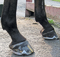
FIGURE 4. Grand Circuit Morrison shoe with a 2-degree wedge demonstrate both placement and the tapered toe.
Wildenstein left the horse in open-heeled shoes to minimize concussion to the back of the foot. He reminded me that the wings of the coffin bone turn from cartilage to bone as horses age. He said a bar shoe would have increased the ground-reaction forces that speed up ossification and would have limited normal hoof expansion and contraction. Decreased circulation behind the quarters would follow with less heel growth.
Lesson #3: Managing a relatively sound, but imperfectly built horse requires attention to age as well as pain or biomechanical problems.
The vets recommended corrective shoeing and offered an impressive arsenal of treatment options. Besides appropriate conditioning, the easiest and cheapest line of defense against OA in performance horses is providing good nutrition along with oral and injectable supplements designed to curb inflammatory reactions and to support healthy joint tissues.
Lobster enjoyed an effective, but minimal, regime of daily MSM (a source of bio-available sulfur to limit inflammatory response) and weekly intramuscular injections of Glucosamine (to support synovial tissues).
Other popular OA treatments include Adequan (polysulfated glycosamineglycan) and Legend (hyaluronate sodium) that are given by intramuscular injection to support the cells in the synovial lining that produces joint fluid.
Various chemicals can also be injected directly into the joints to calm painful inflammatory response and support a healthy synovium. These range from steroids to hyaluronic acid to IRAP. Though they are expensive, temporary and carry a risk of infection, joint injections are commonly used with arthritic horses. Although divided about the long-term benefits, some vets maintain repeated steroid injections hasten the degeneration of joint cartilage.
A last option would have been arthrodesis surgery — fusing the pastern joint by mechanical or chemical means. I decided not to pursue this. I felt it would be unwise to subject an older horse to the long, painful recovery.
A better solution was to shoe this horse so all of his joints could move in the smoothest natural arc allowed by his conformation. The problem was how to correct the mechanical problems that placed the pasterns under too much stress.
Lesson #4: As a rider eager to get back to my summer show schedule, I wanted a quick and logical shoeing solution. However, Wildenstein indicated a quick fix would be short-lived.
With radiographs in hand, he added a 3-degree wedge to the shoe, raising the heel and correcting the angle of the bones inside the hooves and pasterns. The more upright foot was trimmed to accommodate a matched pair of wedge pads.
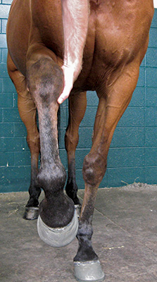
FIGURE 5. While the abaxial deviation in the radius is difficult to see, the offset metacarpal (cannon bone) leaves the knee slightly medially. The fetlock varus stance in both legs comes primarily from a higher up abaxial rotation, partially corrected by an axial deviation below the fetlock that was greater in the right than the left foot.
The solution was temporary indeed. Before the next shoeing, the horse’s conformation and weight took over and his pasterns did not remain at the angle created by the wedges. This extreme solution could only be used for so long.
A steep wedge risked crushing the heels in the long term, but would immediately throw too much weight onto the toe. In fact, the toes became sore while wearing the second set of shoes and we quickly reached the limit of what steep wedges would provide without creating a new problem.
Wildenstein used Sound Horse Technologies’ Morrison Roller Motion Series I (Blue Pad) glue-on shoes for the two shoeings that got us through the end of our summer show season. These padded shoes incorporated a shallower 2-degree wedge.
The glue-ons looked unorthodox for the hunter ring and the trainer felt Lobster moved strangely in them, but they had advantages. The tapered design at the front one-third of the shoe eases breakover, decreasing ground-reaction forces in the last part of the weight-bearing phase of his stride (Figure 3 and 4). A short shoe design from front to back allowed the farrier to set the shoe well back. This helped move the foot’s balance point back to the center of the coffin joint without extending the heels too far under the bulbs.
Shortening the duration of breakover phase of the stride also minimizes the strain out on the soft tissues.
Solving More Than One Problem
The next element of Wildenstein’s shoeing prescription addressed the various pathologies behind the horse’s apparent fetlock varus conformation and anatomical deviations beginning at the elbow.
In both forelimbs, a rotation deviation in the radius indicated an abaxial rotation. Since this horse is carpal valgus, the canon bone descends from the knee slightly medially. From the fetlock on down, he exhibited axial rotations (greater in the right limb) that partially obscure the greater abaxial rotation higher up (Figure 5).
The defects show up as paddling while he moves. Since he toes out appreciably while standing still, positioning radiograph plates for lateral views of the pasterns can be tough.
Six weeks spent in soft aluminum shoes revealed the natural point of breakover is not at the center of his hoof, but rather running lateral to it. Wildenstein says greater axial deviation in the left leg — one that corrects the conformation with respect to the appearance of the leg, but does not change motion as governed by the larger abaxial deviation — caused a crushed medial heel.
Wildenstein says it’s important to look at the whole leg even when thinking about a treatment strategy for a single arthritic joint. As a general rule, the effects of any bony deviation become more severe with a shorter vertical distance when fewer joints are involved.
There was both axial and abaxial rotation distributed over several joints. Even with the pastern’s axial rotations, the coffin joint could take up some of the force created by the defect extending over a short vertical distance. This leads to the need to assess all conformational concerns and effects before applying the appropriate shoes.
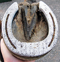
FIGURE 6. This corrective shoeing strategy followed the angle of the pastern axis and matched the shoe to the orientation of the coffin bone rather than the hoof wall. This provided breakover on the lateral side of the toe (right front).
Wildenstein’s shoeing prescription formed an aggressive compromise between an engineering solution to a hyperflexed or broken-back pastern and angular deviation. Therapeutic shoeing goals included raising Lobster’s heel and easing breakover.
After the summer of glue-on shoes, Wildenstein suggested flat shoes for the winter months. The plan was to use an open-heeled shoe to maximize circulation and to trim only as needed to position the hoof behind the quarters.
The long-term options were to prop up the heel without damaging the hoof and coffin bone, and to ease breakover. But by the following spring, there was no increase in heel growth.
With glue-on shoes, Wildenstein felt he could safely use wedge pads indefinitely. However, a wedged aluminum shoe would continue to compress the heels, slow hoof growth and hasten ossification of the coffin bone. A wedge pad made of leather or similar material used with a nail-on shoe would have to be hard enough to keep the clinches stable.
Though many farriers do not share his opinion, Wildenstein feels soft wedge pads are of limited value. Because the coffin bone hangs off the inside of the hoof wall, nailing into it transmits the concussion to this delicate bone and up the leg.
By comparison, glue-on shoes attach the shoe and internal wedge to the entire hoof wall. They can help raise the back of the coffin bone to correct the whole pastern axis while minimizing ground-reaction forces. Thanks to the very soft, black-pad-wedged glue-on shoes, the horse did better than if leather or polyurethane pads had been used with nailed-on steel shoes.
By not being constrained by nail holes or the hoof wall, a farrier can set the glue-on shoe without respect to the hoof’s conformation and place the point of breakover wherever he wishes. In Lobster’s case, the shoe was applied to follow the orientation of the coffin bone — not the hoof capsule. This allowed him to breakover at a point lateral to the center of his toe (Figure 6).
Aggressive Compromises In The Real World
When Lobster was 16, we were both ready to step down from active duty in the show ring. Though progression of front-end arthritis had slowed without resorting to joint injections or low doses of medication, I needed a less elaborate and less expensive shoeing protocol.
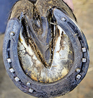
FIGURE 7. A simpler, less expensive winter compromise was a Mustad Equi-Librium steel shoe with ice nails and calks. The left front is still set back to allow speed breakover in any direction.
Since I was looking for a shoeing approach based on nail-on shoes, we abandoned the wedging to focus on easing breakover. Wildenstein used a pair of Mustad Equi-Librium steel shoes (Figure 7). With flat shoes, we’d have to accept the shallow angle with a sloping right front hoof and pastern.
Whether using the thick Equi-Librium shoe or a thinner Eventer shoe, Wildenstein says it’s important to set the shoe well back to place the center of rotation directly below the coffin joint. Even though we wanted to avoid sticking the foot to the ground, we still needed to provide sufficient traction in different environments.
Lobster continued to work and be shown on grass in the Morrison and Roller Motion shoes without a problem. In wet conditions, he would forge since the wide-web shoes reduced traction and allowed the foot to slip backward slightly during breakover. In sand and on harder ground, this problem disappeared. Wildenstein added calks to the toe of the Equi-Librium shoes for winter.
Conclusion
Since OA is best handled by early discovery, careful conditioning and planned corrective shoeing, the farrier plays a unique and substantial role. If farriers can play a part in treating arthritic problems, then they can help owners and trainers recognize why these concerns should to be caught early and managed effectively.
With corrective shoeing, the primary task facing the owner of a middle-aged horse is careful management that involves working within his conformation. The same can be said for younger horses, as shoeing prescriptions vary with the horse’s age, history, environment and job description.


Post a comment
Report Abusive Comment